Unlocking Nature's Pharmacy: X-ray Crystallography Fragment Screening for Natural Product Drug Discovery
This article provides a comprehensive guide to X-ray crystallography fragment screening with natural products, a cutting-edge approach in modern drug discovery.
Unlocking Nature's Pharmacy: X-ray Crystallography Fragment Screening for Natural Product Drug Discovery
Abstract
This article provides a comprehensive guide to X-ray crystallography fragment screening with natural products, a cutting-edge approach in modern drug discovery. Aimed at researchers and drug development professionals, it explores the unique chemical space of natural products as fragment libraries, details the experimental workflow from library preparation to structure determination, addresses key technical challenges, and validates the method's superiority against other screening techniques. We synthesize current best practices and future directions for integrating this powerful methodology into the next generation of therapeutics.
Why Natural Products? The Unique Power of Nature's Fragments for Targeted Screening
The Renaissance of Natural Products in Modern Drug Discovery
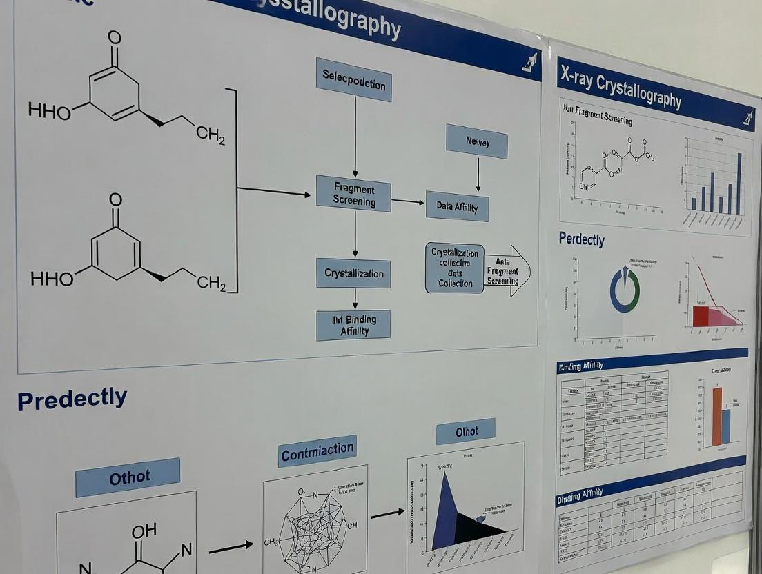
Application Notes
The integration of natural products (NPs) into fragment-based drug discovery (FBDD) pipelines, underpinned by X-ray crystallography, addresses historical limitations of NP drug discovery—namely, complexity, derivatization challenges, and target deconvolution. Modern strategies treat NPs as privileged fragment libraries, leveraging their inherent structural complexity and biomolecular recognition properties.
Note 1: NP-Focused Fragment Libraries. Curated libraries like the NIH Natural Product Integrity (NPI) library (~1,000 compounds) and commercial collections (~5,000 pre-fractionated extracts) are screened using biophysical methods. Surface Plasmon Resonance (SPR) and Microscale Thermophoresis (MST) provide primary hit identification, with X-ray crystallography serving as the definitive structural validation tool. This tandem approach reduces false positives from promiscuous binders common in crude extracts.
Note 2: Synergy with Genomics and Metabolomics. The resurgence is fueled by genomic mining and metabolomics, enabling targeted isolation of NPs from microbial sources. For example, genome sequencing of Streptomyces species reveals cryptic biosynthetic gene clusters, increasing the probability of discovering novel chemotypes with unique binding modalities.
Note 3: Targeting "Undruggable" Pockets. NPs, with their complex three-dimensional scaffolds, are particularly effective at binding to shallow protein-protein interaction (PPI) interfaces and allosteric sites often considered "undruggable" by synthetic flat molecules. X-ray crystallography fragment screening of NPs has yielded hits against targets like KRAS and Myc.
Quantitative Data Summary
Table 1: Recent NP-Derived Drug Approvals (2020-2024)
| NP Source | Drug Name (Approval Year) | Target/Indication | Discovery Approach |
|---|---|---|---|
| Marine Bacterium | Lurbinectedin (2020) | DNA minor groove, SCLC | NP analog synthesis |
| Plant (Artemisia annua) | Artemisinin-based combos (WHO rec.) | Malaria | NP derivatization |
| Fungus | Fosmanogepix (Phase III) | GPI-anchored proteins, Fungal infection | NP-inspired synthetic |
| Synthetic Biology (Yeast) | Hyaluronic acid variants (2023) | Dermal fillers | Biosynthetic engineering |
Table 2: Comparison of Screening Methods for NP Fragment Screening
| Method | Throughput | Sample Required | Kd Range | Key Advantage for NPs |
|---|---|---|---|---|
| X-ray Crystallography | Low | ~100-500 µM, 5-10 mg/mL | mM to µM | Direct 3D binding mode visualization |
| SPR | Medium-High | ~1-100 µM | nM to mM | Label-free, kinetic data |
| MST | Medium | ~nM-µM | pM to mM | Works in complex buffers (e.g., crude extract) |
| NMR (Ligand-observed) | Low-Medium | ~10-500 µM | µM to mM | Detects weak binders, identifies binding moiety |
Experimental Protocols
Protocol 1: X-ray Crystallography Fragment Screening of a Pre-fractionated Natural Product Library
Objective: To identify and characterize fragments from a pre-fractionated NP library binding to a purified protein target.
Materials:
- Purified, crystallizable protein target (>95% purity, 10 mg/mL).
- Pre-fractionated NP library (e.g., 384-well plate, 20 mM in DMSO).
- Crystallization reagents and plates.
- Synchrotron or home-source X-ray diffractometer.
Procedure:
- Co-crystallization Soaking: a. Grow native protein crystals using established vapor diffusion methods. b. Prepare soaking solutions: Add 0.5 µL of NP library compound (20 mM in DMSO) to 49.5 µL of reservoir solution to create a 0.2 mM soaking solution (1% DMSO). c. Transfer a single native crystal into 10 µL of soaking solution. Incubate for 2-24 hours at the crystallization temperature. d. Cryo-protect the crystal and flash-cool in liquid nitrogen.
Data Collection & Processing: a. Collect a complete X-ray diffraction dataset for each soaked crystal at a synchrotron beamline. b. Process data using XDS or DIALS. Scale with AIMLESS. c. Solve the structure by molecular replacement using the apo protein model (Phaser). d. Refine the structure iteratively with REFMAC5 or phenix.refine.
Ligand Fitting & Analysis: a. Examine difference electron density maps (Fo-Fc, contoured at +3.0 σ) for positive density in the protein's active site or pockets. b. For clear density, fit the NP fragment using Coot. Use the ELBOW program to generate geometry restraints. c. Validate the binding pose: Analyze interactions (H-bonds, hydrophobic contacts) and ligand geometry.
Protocol 2: Activity-Guided Fractionation Coupled with SPR Primary Screening
Objective: To isolate and identify active compounds from a crude natural extract.
Materials:
- Crude natural extract (e.g., plant, marine sponge).
- SPR system (e.g., Biacore, Sierra Sensors) with immobilized target protein.
- HPLC system with fraction collector.
- Analytical LC-MS.
Procedure:
- SPR Primary Screen: a. Immobilize purified target protein on a CM5 sensor chip via amine coupling. a. Inject crude extract (diluted in running buffer to 0.1 mg/mL) over the target and reference flow cells at 30 µL/min. b. Identify "hit" extracts that show a concentration-dependent binding response (Response Units, RU).
Bioassay-Guided Fractionation: a. Fractionate the "hit" crude extract using reverse-phase flash chromatography. b. Collect 96 fractions in a deep-well plate. c. Dry down fractions and resuspend in DMSO for secondary SPR screening. d. Screen all fractions against the immobilized target via SPR. Pool active fractions.
Iterative Purification & Identification: a. Subject active pools to semi-preparative HPLC for further separation. b. Repeat SPR screening of sub-fractions until pure active compounds are obtained. c. Determine structure of active pure compound using NMR and High-Resolution Mass Spectrometry (HR-MS).
Diagrams
Title: NP Fragment Screening & Optimization Workflow
Title: NP Fragment Allosteric PPI Inhibition
The Scientist's Toolkit
Table 3: Key Research Reagent Solutions for NP X-ray Crystallography Screening
| Item | Function & Rationale |
|---|---|
| Prefractionated NP Library (e.g., Selleckchem Natural Product Library) | Minimizes complexity for screening; provides semi-pure compounds in standardized formats (96/384-well plates), reducing interference in crystallography and biophysical assays. |
| Crystallization Screening Kits (e.g., Morpheus HT-96, MemGold2) | Broad-spectrum sparse matrix screens optimized for membrane proteins and protein-ligand complexes, increasing co-crystallization success rates with NP fragments. |
| SPR Sensor Chips (Series S, CM5) | Gold standard for label-free, real-time binding kinetics. CM5 chips allow stable immobilization of diverse protein targets via amine coupling for primary NP extract screening. |
| Microscale Thermophoresis (MST) Capillaries | Enables binding affinity measurement in solution with minimal sample consumption. Ideal for screening crude extracts or unstable proteins unsuitable for crystallization. |
| Cryoprotectant Solutions (e.g., Paratone-N, LV Oil) | Protects crystals during flash-cooling for data collection at cryogenic temperatures, preventing ice formation that degrades diffraction quality. |
| Ligand Restraint Generation Software (e.g., GRADE, eLBOW) | Automates the creation of accurate geometry and energy restraints for novel, complex NP ligands, which are often absent from standard libraries, for refinement in X-ray structures. |
| HEPES Buffered Saline (HBS-EP+) for SPR | Standard running buffer for SPR assays; low non-specific binding and compatible with DMSO from compound stocks, ensuring stable baselines during NP library injections. |
Application Notes
Fragment-based drug discovery (FBDD) is a core methodology in modern lead generation, where libraries of low molecular weight compounds (<300 Da) are screened to identify weak binders to a target protein. These fragments are subsequently elaborated into potent leads. The composition of the fragment library is critical. This document contrasts two primary sources: natural products and synthetic libraries, with a focus on their application in X-ray crystallographic screening for novel pharmacophore discovery.
Natural Product Fragments: Derived from biologically pre-validated scaffolds, natural product fragments (NPFs) offer high structural diversity and three-dimensional complexity, often rich in sp3-hybridized carbons and stereocenters. They sample chemical space evolved for biomolecular interaction, potentially leading to higher hit rates for challenging targets and improved developability. However, their supply, structural complexity (which can hinder synthetic optimization), and potential for nuisance compounds (e.g., pan-assay interference compounds) require careful curation.
Synthetic (Rule-based) Fragment Libraries: Designed using rules like the "Rule of Three," these libraries prioritize synthetic tractability, solubility, and purity. They ensure chemical space is covered efficiently and facilitate rapid, straightforward hit-to-lead chemistry. The primary risk is a potential over-reliance on flat, aromatic scaffolds, leading to less novel and more lipophilic leads.
A hybrid approach, integrating a subset of NPFs into a standard synthetic library, is increasingly adopted to balance novelty with practicality.
Table 1: Quantitative Comparison of Fragment Sources
| Parameter | Synthetic Fragment Libraries (Standard) | Natural Product-Derived Fragments (Curated) | Notes |
|---|---|---|---|
| Avg. Molecular Weight (Da) | 150 - 250 | 200 - 300 | NPFs are slightly heavier but within fragment range. |
| Avg. Heavy Atom Count | 10 - 18 | 14 - 22 | Reflects greater complexity. |
| Avg. Fraction sp3 (Fsp3) | 0.25 - 0.40 | 0.45 - 0.70 | Key metric for 3D shape. |
| Avg. Calculated LogP (cLogP) | 0.5 - 2.5 | 0.0 - 3.0 | Wider distribution for NPs. |
| Typical Solubility (mM) | >1.0 (in aqueous buffer) | Variable, often >0.5 | Requires pre-screening for NPs. |
| Structural Diversity | Moderate (rule-guided) | High (biosynthesis-driven) | NPs access underrepresented chemotypes. |
| Synthetic Tractability | High | Moderate to Low | NP elaboration can be challenging. |
| Typical X-ray Screening Hit Rate | 1 - 5% | 3 - 10% (for compatible targets) | NPs show elevated rates for some target classes. |
Experimental Protocols
Protocol 1: Preparation of a Hybrid Fragment Library for Crystallographic Screening Objective: To create a 500-compound screening library comprising 400 synthetic fragments and 100 natural product fragments.
Materials: See "The Scientist's Toolkit" (Table 2).
Procedure:
- Library Design:
- Select 400 synthetic fragments from commercial sources adhering to Rule of Three (MW ≤ 300, cLogP ≤ 3, HBD ≤ 3, HBA ≤ 3, PSA ≤ 60 Ų). Enforce >85% purity.
- Select 100 natural product-derived fragments from vendors specializing in NP-like scaffolds. Apply filters: MW ≤ 300, cLogP ≤ 3.5, heavy atoms ≤ 22, and pan-assay interference compound (PAINS) alerts removed. Confirm purity >90%.
- Perform computational diversity analysis (e.g., using Tanimoto similarity on extended connectivity fingerprints) to ensure minimal overlap between the two subsets.
- Stock Solution Preparation:
- Prepare 100 mM stock solutions of all fragments in 100% DMSO. Use a calibrated acoustic dispenser for accuracy.
- For NPFs with suspected solubility issues, confirm stock solubility by visual inspection and nephelometry.
- Crystallography Screening Cocktail Formulation:
- Use software (e.g., Cocktail Wizard) to group 4-8 fragments per cocktail based on complementary shapes and sizes to avoid crystal packing interference.
- Prepare each cocktail by mixing equimolar volumes of individual stocks to achieve a final per-fragment concentration of 25-50 mM in 100% DMSO.
- Critical: For cocktails containing NPFs, include one "NPF-only" cocktail per plate to monitor for unique diffraction artifacts.
- Protein Co-Crystallization/Soaking:
- Co-crystallization: Mix protein solution (at 5-10 mg/mL) with screening cocktail at a 9:1 ratio (v/v), yielding a final per-fragment concentration of 2.5-5 mM and 10% DMSO. Set up crystallization trays immediately.
- Soaking: Transfer pre-grown crystals to a stabilizing solution containing 5% (v/v) of the screening cocktail (final per-fragment concentration ~1-2 mM). Soak for 2-24 hours.
- Data Collection & Analysis:
- Flash-cool crystals in liquid nitrogen. Collect X-ray diffraction data to a resolution of ≤2.2 Å.
- Process data with standard software (e.g., XDS, autoPROC).
- Solve structures by molecular replacement. Use automated software (e.g., PanDDA, FragMAX) to identify electron density outliers consistent with bound fragments.
Protocol 2: Validation and Hit Elaboration of a Natural Product Fragment Hit Objective: To validate a fragment hit from an NPF library and design analogs for synthetic elaboration.
Procedure:
- Orthogonal Biophysical Validation:
- Surface Plasmon Resonance (SPR): Test the pure NPF hit in a dose-response series (0.5 μM to 500 μM) to confirm binding affinity (KD) and obtain kinetic parameters.
- Ligand-Observed NMR: Perform ( ^1H ) CPMG or STD-NMR experiments to confirm binding in solution.
- Medicinal Chemistry Analysis:
- Using the crystal structure, identify key interactions (hydrogen bonds, hydrophobic contacts, water bridges).
- Define the fragment's growth vectors (atoms/groups amenable to chemical modification without clashing with the protein).
- Analog Sourcing and Testing:
- Search commercial and in-house compound collections for analogs of the NPF core (e.g., simplified synthetic versions, natural analogs).
- Screen these analogs using the crystallographic soaking protocol to establish a preliminary structure-activity relationship.
- Design of Elaborated Compounds:
- Use structure-based design software (e.g., SeeSAR, MOE) to propose chemical elaborations along the growth vectors.
- Prioritize synthetically accessible designs that enhance key interactions.
Visualizations
FBDD Workflow Integrating NP and Synthetic Fragments
Characteristics Driving Hybrid Library Design
The Scientist's Toolkit
Table 2: Essential Research Reagents & Materials
| Item/Reagent | Function in Protocol | Key Consideration |
|---|---|---|
| Commercially Available Fragment Libraries | Source of pre-plated, characterized synthetic fragments. | Ensure compliance with chosen rules (e.g., Rule of 3) and availability of chemical matter for follow-up. |
| Natural Product Fragment Collections | Source of sp3-rich, complex scaffolds (e.g., from AnalytiCon, TimTec NP Library). | Requires stringent PAINS filtering and solubility validation prior to screening. |
| DMSO (High-Purity, Anhydrous) | Universal solvent for fragment stock solutions. | Low water content is critical to prevent crystal degradation during soaking. |
| Acoustic Liquid Dispenser (e.g., Echo) | Non-contact, precise transfer of nanoliter volumes of DMSO stocks. | Enables accurate cocktail formulation and minimizes DMSO volume in assays. |
| Crystallization Plates (SBS Format) | For setting up protein-co-crystal trials with fragment cocktails. | Compatibility with automated imaging systems is essential for high-throughput. |
| Cocktail Design Software (e.g., Cocktail Wizard, SILVER) | Groups fragments into cocktails to maximize success in crystallographic screening. | Algorithms must avoid combining fragments that could sterically clash in the binding site. |
| Pan-Dataset Density Analysis (PanDDA) Software | Statistical method to identify weak fragment binding in large crystallographic datasets. | Crucial for detecting low-occupancy, high-solubility fragment hits, common with NPFs. |
| Biophysical Validation Suite (SPR, NMR) | Orthogonal validation of crystallographic hits to rule out false positives. | SPR requires careful immobilization of the target protein; NMR requires isotopic labeling for protein-observed methods. |
Natural products (NPs) and their derivatives represent a pre-validated source of bioactive chemical matter, refined by evolution to interact with biological macromolecules. Within X-ray crystallography-based fragment screening (XCFS) campaigns, NP-derived fragment libraries offer distinct advantages over synthetic libraries by providing:
- Enhanced Complexity and 3D Shape: NPs often exhibit greater stereochemical complexity and sp³-hybridized character, leading to more globular, protein-surface-compliant shapes. This improves the likelihood of high-affinity, selective binding.
- High Skeletal Diversity: Biosynthetic pathways generate core scaffolds (e.g., polyketides, alkaloids, terpenoids) that are largely underrepresented in synthetic medicinal chemistry, accessing novel regions of chemical space.
- Evolved Bioactivity: NPs are biosynthesized for ecological purpose, meaning they possess innate, optimized bioactivity and "drug-likeness." This evolutionary pre-selection increases hit rates for biologically relevant targets.
Integrating NP fragments into XCFS accelerates the identification of novel, high-quality binding motifs for challenging drug targets, such as protein-protein interactions and allosteric sites.
Table 1: Comparison of Fragment Library Properties
| Property | Synthetic Fragment Library (Typical) | Natural Product-Derived Fragment Library (Typical) | Data Source / Reference |
|---|---|---|---|
| Avg. Molecular Weight (Da) | 180-250 | 200-320 | Analysis of commercial libraries (ZINC, COCONUT DB) |
| Avg. Fraction sp³ Carbons (Fsp³) | 0.25-0.35 | 0.40-0.70 | Analysis of commercial libraries (ZINC, COCONUT DB) |
| Avg. Number of Stereocenters | 0-1 | 2-5 | Analysis of commercial libraries (ZINC, COCONUT DB) |
| Represented Scaffold Classes | Aromatic heterocycles, simple aliphatics | Polyketides, alkaloids, terpenoids, flavonoids | Chemical taxonomy studies |
| XCFS Hit Rate (Range) | 0.5% - 5% | 2% - 10% (for compatible targets) | Published XCFS campaigns (e.g., COVID Moonshot) |
| LE (Ligand Efficiency) Avg. | 0.30-0.45 | 0.35-0.50 | Retrospective fragment screening analyses |
Table 2: Exemplar NP Fragments from X-ray Screening Campaigns
| NP Fragment Core | Source Organism | Target Protein | PDB ID (Example) | Binding Mode Key Feature |
|---|---|---|---|---|
| Adenosine derivative | Marine sponge | SARS-CoV-2 Mpro | 7LC5 | Binds proximal to catalytic dyad |
| Indole alkaloid core | Streptomyces sp. | Beta-Lactamase | 6U88 | Occupies oxyanion pocket |
| Diterpene fragment | Plant extract | KRAS G12C | 8SPY | Engages cryptic allosteric pocket |
Experimental Protocols
Protocol 1: Construction of a NP-Derived Fragment Library for XCFS
Objective: To curate and prepare a diverse, X-ray crystallography-compliant library of fragments derived from natural products.
Materials: See "The Scientist's Toolkit" (Section 5). Procedure:
- Database Mining: Query natural product databases (e.g., COCONUT, NPASS) with filters: Molecular Weight ≤ 300 Da, Rotatable Bonds ≤ 5, XLogP ≤ 3.2.
- Chemical Clustering: Perform scaffold analysis (e.g., using RDKit). Cluster compounds by Bemis-Murcko frameworks. Select representatives from each major NP scaffold class to maximize diversity.
- Property Filtering: Apply "Rule of 3" filters and remove pan-assay interference compounds (PAINS) using dedicated filter sets.
- Commercial Sourcing & Validation: Purchase selected compounds. Confirm purity (>95% by LC-MS) and identity (NMR). Prepare 100 mM stock solutions in DMSO.
- Crystallography Suite Preparation: Prepare a master mix of fragments by combining stocks to a final concentration of 5-10 mM each in a compatible cryoprotectant buffer (e.g., 25% PEG 400, 75% mother liquor). Final DMSO concentration should be <5%.
- Soaking Solution Prep: For each fragment cocktail (typically 4-8 fragments), combine 1 µL of the master mix with 9 µL of crystal stabilization buffer immediately prior to soaking.
Protocol 2: X-ray Crystallographic Fragment Screening via Soaking
Objective: To screen a NP fragment library against a target protein crystal to identify bound fragments.
Materials: Pre-formed protein crystals, NP fragment cocktails, crystallization plates, synchrotron or home X-ray source. Procedure:
- Crystal Preparation: Harvest a single, well-diffracting protein crystal into a drop of mother liquor.
- Fragment Soaking: Transfer the crystal into a 1 µL drop of the prepared fragment cocktail solution. Incubate for 30 minutes to 2 hours at the crystallization temperature.
- Cryo-Cooling: After soaking, swiftly loop the crystal and cryo-cool it in liquid nitrogen.
- Data Collection: Collect a complete X-ray diffraction dataset at a synchrotron beamline (e.g., 100K temperature, high flux).
- Data Processing: Index, integrate, and scale the data (using XDS, autoPROC, or DIALS).
- Difference Map Analysis: Refine the protein model against the data, then compute an mFo-DFc ("difference") electron density map contoured at +3.0 σ. Visually inspect for unambiguous positive density adjacent to the protein, indicating a bound fragment.
- Fragment Modeling & Refinement: Fit an appropriate fragment structure into the electron density and perform iterative cycles of refinement (e.g., with REFMAC5 or phenix.refine).
Visualizations
Diagram Title: XCFS Workflow with NP Fragments
Diagram Title: NP Fragment to Lead Progression
The Scientist's Toolkit
Table 3: Key Research Reagent Solutions for NP Fragment XCFS
| Item | Function in NP Fragment XCFS |
|---|---|
| COCONUT / NPASS Database | Primary sources for natural product structures and bioactivity data for virtual library construction. |
| Rule of 3 Filtering Software (e.g., RDKit) | Computationally filters large NP datasets into fragment-like chemical space (MW <300, HBD ≤3, etc.). |
| PAINS / REOS Filter Sets | Identifies and removes compounds with undesirable functional groups prone to assay interference or reactivity. |
| DMSO-d₆ for NMR | Solvent for confirming compound identity and purity of sourced NP fragments prior to screening. |
| Crystallization Screen Kits (e.g., Morpheus, JCGSG) | For initial crystallization and optimization of the target protein to obtain robust, diffraction-quality crystals. |
| Synchrotron Beamtime | Essential high-intensity X-ray source for rapid, high-throughput collection of fragment-screened crystal datasets. |
| Coot Molecular Graphics | Software for visual inspection of electron density maps and manual modeling/refitting of bound fragments. |
| PanDDA (Pan-Dataset Density Analysis) | Computational method to identify weak, low-occupancy fragment binding events across multiple datasets. |
Historical Success Stories and the Case for Fragment-Based Approaches
Within the broader thesis on advancing natural products research through X-ray crystallography fragment screening, this document details key historical successes and provides standardized protocols. Fragment-based drug discovery (FBDD) has proven particularly powerful in targeting challenging protein sites, with several drugs now on the market originating from this approach.
Table 1: Marketed Drugs Originating from Fragment-Based Approaches
| Drug Name (Brand) | Target | Indication | Approx. Fragment MW (Da) | Final Drug MW (Da) | Year Approved | Key Technique for Screening |
|---|---|---|---|---|---|---|
| Vemurafenib (Zelboraf) | B-Raf V600E mutant kinase | Melanoma | ~230 | 489.9 | 2011 | X-ray Crystallography |
| Venetoclax (Venclexta) | BCL-2 | CLL, AML | ~250 | 868.4 | 2016 | NMR, X-ray Crystallography |
| Sotorasib (Lumakras) | KRAS G12C | NSCLC | ~150 | 560.7 | 2021 | X-ray Crystallography |
| Pexidartinib (Turalio) | CSF1R, KIT | Tenosynovial Giant Cell Tumor | ~200 | 529.5 | 2019 | Biochemical Screening, X-ray |
Table 2: Key Metrics from Natural Product Fragment Screening Campaigns (Representative)
| Campaign Target (Class) | Library Size | Hit Rate (%) | Avg. Fragment Ligand Efficiency (LE) | Best LE | Structure Elucidated Via |
|---|---|---|---|---|---|
| SARS-CoV-2 Mpro | 1,213 | 3.5 | 0.32 | 0.41 | MicroED, X-ray |
| β-Lactamase (Antibiotic Resistance) | 500 (NP-inspired) | 5.2 | 0.35 | 0.48 | X-ray Crystallography |
| Hsp70 (Oncology) | ~800 | 2.1 | 0.28 | 0.39 | X-ray & NMR |
Application Notes & Detailed Protocols
Protocol 1: Library Design & Preparation for NP-Inspired Fragment Screening
Objective: To construct a fragment library enriched with natural product-like scaffolds for primary X-ray crystallographic screening. Materials: See "The Scientist's Toolkit" below. Procedure:
- Virtual Library Curation:
- Source 3D structures of diverse natural products from databases (e.g., COCONUT, ZINC Natural Products).
- Apply in silico fragmentation using retrosynthetic combinatorial analysis procedure (RECAP) rules to generate plausible fragment scaffolds.
- Filter fragments using "Rule of 3" (MW ≤ 300, cLogP ≤ 3, HBD ≤ 3, HBA ≤ 3, rotatable bonds ≤ 3). Allow modest latitude for ring complexity.
- Physical Library Assembly:
- Procure or synthesize selected fragments (purity ≥95% by HPLC/LCMS).
- Prepare 100 mM stock solutions in DMSO-d6 for initial NMR validation.
- Dilute to 1 M in DMSO for crystallography screening stocks. Store at -20°C under desiccant.
- Pre-Screen Validation:
- Confirm solubility in aqueous buffer (PBS, pH 7.4) at final screening concentration (typically 10-50 mM) using nephelometry.
- Confirm lack of aggregation via 1D 1H NMR in buffer.
Protocol 2: High-Throughput X-ray Crystallography Fragment Soaking
Objective: To screen a fragment library against a pre-formed protein crystal to identify bound ligands. Materials: Purified target protein (>95%), crystallization reagents, fragment library plates, micro-sized loops. Procedure:
- Protein Crystallization & Harvest:
- Grow crystals of the target protein using optimized vapor-diffusion conditions.
- Harvest crystals using a micro-loop and briefly transfer to a stabilizing/cryo-protection solution.
- Fragment Soaking:
- Prepare soaking solution: Add fragment from screening stock to crystallization mother liquor to achieve a final concentration of 50-100 mM. Include 5% (v/v) DMSO as a carrier control.
- Transfer a single crystal into 2 µL of soaking solution. Incubate for 30 minutes to 24 hours in a humidity chamber (time determined empirically).
- Cryo-Cooling & Data Collection:
- After soaking, quickly loop the crystal and plunge into liquid nitrogen.
- Collect X-ray diffraction data at a synchrotron or home-source (wavelength ~1.0 Å). Aim for resolution ≤2.2 Å.
- Data Processing & Analysis:
- Process data (index, integrate, scale) using software like XDS, DIALS, or HKL-3000.
- Solve structures by molecular replacement using the apo-protein model.
- Calculate initial |Fo| - |Fc| difference maps (omit maps) to identify positive electron density peaks indicative of bound fragments.
- Model fragments into density using Coot and refine with REFMAC5 or phenix.refine.
Protocol 3: Hit Validation & Progression via SPR & Ligand-Observed NMR
Objective: To validate crystallographic hits and determine initial binding metrics. Procedure: A. Surface Plasmon Resonance (SPR):
- Immobilize target protein on a CMS chip via amine coupling to achieve ~5000-10000 RU response.
- Perform single-cycle kinetics: Inject a series of fragment concentrations (0.5-1000 µM) in HBS-EP+ buffer (containing 2% DMSO) at a flow rate of 30 µL/min.
- Analyze sensorgrams using a 1:1 binding model to extract KD, kon, and koff. A confirmed hit typically has KD < 1 mM.
B. Ligand-Observed NMR (CPMG & Water-LOGSY):
- Prepare sample: 20 µM protein in PBS, 50 µM fragment (2.5:1 ratio), 10% D2O, 0.02% AZR.
- For CPMG: Collect 1D 1H spectra with a T2 filter (Δ = 400 ms) to attenuate protein signals. Signal attenuation of fragment peaks indicates binding.
- For Water-LOGSY: Use excitation sculpting to suppress water. Invert magnetization transfer from water to bound ligand. Negative NOE peaks for the fragment indicate binding.
- Compare spectra with protein-free controls.
Visualizations
The Scientist's Toolkit
Table 3: Essential Research Reagents & Materials for FBDD with X-ray Crystallography
| Item | Function in Workflow | Example/Supplier Notes |
|---|---|---|
| High-Purity Target Protein (>95%) | Essential for forming diffraction-quality crystals. | Recombinant expression systems (E. coli, insect cells). Purification tags (His, GST). |
| Fragment Library (NP-focused) | Source of chemical starting points. | Commercially available (e.g., Enamine REAL Space) or custom-curated. 500-2000 compounds. |
| Crystallization Kits | Initial screening of crystallization conditions. | Sparse-matrix screens (e.g., Hampton Research Index, MD kits). |
| Micro Crystallography Loops | Harvesting and mounting fragile crystals. | MiTeGen loops in various sizes (50-200 µm). |
| Liquid Nitrogen Dewar | Cryo-cooling crystals for data collection. | For storage and transport. |
| Synchrotron Beam Time | High-intensity X-ray source for data collection. | Facilities: APS, ESRF, Diamond Light Source. |
| DMSO-d6 | Solvent for fragment stocks and NMR validation. | Anhydrous, 99.9% atom D. |
| SPR Chip (CM5) | Immobilization of protein for biophysical validation. | Gold surface with carboxymethylated dextran matrix. |
| NMR Tubes | For ligand-observed NMR binding assays. | 3 mm or 5 mm matched tubes (e.g., Wilmad). |
| Data Processing Software | Turning diffraction images into electron density maps. | XDS, CCP4, Phenix, HKL-3000 suites. |
| Molecular Graphics Software | Visualizing and modeling fragments into density. | Coot, PyMOL, ChimeraX. |
Within the broader thesis on advancing X-ray crystallography fragment screening for novel drug discovery, this application note details the integration of natural product (NP) fragments. NPs are privileged starting points due to their inherent structural complexity, bio-relevance, and proven track record. This document outlines current sourcing strategies and provides protocols for the construction and screening of NP fragment libraries, emphasizing compatibility with high-throughput X-ray crystallography pipelines.
Current Trends in NP Fragment Library Sourcing
The field is moving beyond traditional whole-molecule NP screening towards rational fragmentation and curated library design. Key trends include:
- From Complex NPs to Fragments: Intentional degradation or de novo synthesis of fragments derived from NP cores (e.g., indoles, flavones, pyrans, tropanes) to maintain "NP-likeness" while achieving fragment-like properties (MW <300, cLogP <3).
- Diversity-Oriented Synthesis (DOS): Using NP-inspired scaffolds to generate structurally diverse, three-dimensional fragment libraries with enhanced stereochemical and skeletal complexity compared to synthetic flat aromatics.
- Commercial Availability: Rise of specialized vendors offering pre-plated, X-ray screening-ready NP fragment libraries.
- In Silico Pre-Screening: Computational filtering of virtual NP fragment spaces for desirable physicochemical properties and predicted solubility, crucial for crystallography experiments.
- Focused Libraries: Libraries built around specific NP pharmacophores (e.g., benzopyran, macrocyclic derivatives) to target particular protein families.
Table 1: Selected Commercial and Public Sources for NP Fragment Libraries (Representative Data)
| Source / Library Name | Type of Collection | Approx. Size | Avg. MW (Da) | Avg. cLogP | Key Features / Source | Format for Screening |
|---|---|---|---|---|---|---|
| Enamine REAL Fragment Set (NP Subset) | Synthetic, NP-inspired | ~2,000 | 215 | 1.8 | Designed around NP scaffolds; high Fsp³. | DMSO stock, 96/384-well plates. |
| Life Chemicals NP Fragment Library | Curated commercial | ~1,500 | 220 | 1.9 | Derived from known NP structures; includes alkaloids, terpenoids. | DMSO solution, dry powder. |
| AnalytiCon MEGx NP Fragments | Physically purified NPs & derivatives | ~1,000 | 240 | 2.1 | Direct fragments from microbial/fungal extracts. | Pre-plated in DMSO. |
| NCI Natural Product Set III | Publicly available | ~1,000 | N/A | N/A | Crude and semi-pure natural products, some fragment-like. | Solid samples, requires solubilization. |
| In-house DOS Library (Thesis Work) | Custom synthesized | ~500 | 205 | 1.5 | Synthesized around stereochemically rich NP cores (e.g., decalins). | 100mM in DMSO, 96-well plates. |
Application Notes & Protocols
Protocol 1: Design and Curation of an In-House NP Fragment Library for X-ray Crystallography
Objective: To assemble a 500-member NP fragment library suitable for high-throughput soaking experiments with protein crystals.
Research Reagent Solutions & Essential Materials:
| Item | Function / Explanation |
|---|---|
| DMSO-d6, 99.9% | NMR solvent for compound validation and concentration determination. |
| HPLC-MS Grade DMSO | High-purity, anhydrous DMSO for preparing fragment stock solutions to prevent crystal damage. |
| 96-Well Polypropylene Storage Plates (Sealing) | Chemically resistant plates for long-term storage of 100mM fragment stocks at -80°C. |
| LC-MS System (with PDA/ELSD) | For purity assessment (>95% pure) of all library members. |
| NMR Spectrometer (400 MHz) | For final structural confirmation of synthesized or purchased fragments. |
| Echo 555 Liquid Handler | Non-contact dispenser for precise transfer of nanoliter volumes of fragments into crystal soaking drops. |
Methodology:
- Virtual Library Generation & Filtering:
- Select NP scaffold cores from databases (e.g., COCONUT, LOTUS).
- Generate virtual fragments via in silico retrosynthetic cleavage or purchase available building blocks.
- Filter using rules: MW ≤ 250, cLogP ≤ 2.5, number of heavy atoms 10-18, H-bond donors/acceptors ≤ 5, rotatable bonds ≤ 5. Prioritize fragments with Fsp³ > 0.4.
- Acquisition & Synthesis: Procure commercially available fragments or synthesize via DOS routes to ensure scaffold diversity.
- Quality Control (QC):
- Analyze each compound by UPLC-MS for purity (≥95%).
- Confirm identity and concentration by ¹H NMR in DMSO-d6.
- Assess solubility in aqueous buffer (e.g., 50 mM HEPES pH 7.5) via nephelometry; exclude compounds with >10% precipitation at 10 mM.
- Library Formatting:
- Prepare a master stock plate: dissolve each qualified fragment in HPLC-grade DMSO to a final concentration of 100 mM.
- Seal the plate and store at -80°C.
- Create a daughter plate (10 mM in DMSO) for daily screening use via acoustic dispensing.
Protocol 2: High-Throughput X-ray Crystallography Soaking Experiment with an NP Fragment Library
Objective: To screen a formatted NP fragment library against a target protein crystal via soaking and collect diffraction data.
Methodology:
- Protein Crystal Preparation:
- Grow target protein crystals using optimized vapor diffusion conditions.
- Harvest crystals into a stabilization solution (mother liquor with potential cryoprotectant).
- Fragment Soaking:
- Using an Echo liquid handler, transfer 30 nL of the 10 mM fragment stock (from Protocol 1 daughter plate) into a 96-well sitting drop plate.
- Add 70 nL of crystal stabilization solution to the same well, creating a 100 nL drop with a final fragment concentration of 3 mM.
- Using a loop, transfer a single crystal into the drop. Seal the plate.
- Soak crystals for 30-90 minutes at the crystallization temperature.
- Cryo-Cooling and Data Collection:
- After soaking, quickly loop the crystal and plunge into liquid nitrogen.
- Mount crystals on an automated sample changer.
- Collect X-ray diffraction data at a synchrotron or home source (e.g., 1.0 Å wavelength, 360° rotation, 0.1° oscillation).
- Data Processing & Analysis:
- Process data (index, integrate, scale) with Dials or XDS.
- Use molecular replacement with the apo protein model.
- Perform difference Fourier analysis (|Fo| - |Fc|, φc) to identify electron density for bound fragments.
- Refine fragment-positive hits using REFMAC5 or phenix.refine.
Mandatory Visualizations
Diagram 1: Workflow for Creating an NP Fragment Library
Diagram 2: Crystal Soaking for Fragment Screening
From Compound to Structure: A Step-by-Step Guide to X-ray Fragment Screening Workflows
Application Notes for Natural Product Fragment Screening in X-ray Crystallography
Within the broader thesis of advancing natural product (NP) drug discovery via X-ray crystallography-based fragment screening, the design and preparation of a high-quality screening library is the critical first step. This process bridges the gap between complex NP extracts and structured atomic-level binding data.
Core Challenges & Strategies:
- Sourcing: Moving beyond traditional crude extracts to semi-purified or pure NP fractions to reduce complexity and facilitate deconvolution.
- Solubility: NPs often exhibit poor aqueous solubility. Systematic formulation is required to achieve mM stock concentrations in crystallography-compatible buffers without precipitation.
- Cocktail Formulation: Intelligently mixing multiple fragments into cocktails maximizes beamtime efficiency but requires careful attention to compound compatibility and crystallographic deconvolution.
Key Quantitative Parameters for Library Design The following table summarizes target specifications for an NP-enriched fragment library suitable for X-ray crystallography screening.
Table 1: Target Specifications for an X-ray Crystallography NP Fragment Library
| Parameter | Target Specification | Rationale |
|---|---|---|
| Library Size | 500 – 1000 compounds | Manages complexity while providing sufficient chemical diversity for initial screening. |
| Molecular Weight | ≤ 300 Da | Adheres to Rule of 3 for fragments, promoting efficient binding to discrete pockets. |
| Heavy Atom Count | ≤ 22 | Correlates with smaller size, suitable for fragment-sized binding events. |
| LogP | ≤ 3 | Balances solubility and membrane permeability; lower LogP favors crystallography buffer solubility. |
| Aqueous Solubility | ≥ 1 mM in assay buffer | Essential for achieving saturating concentrations in crystallization drops. |
| Compound Purity | ≥ 90% (by HPLC) | Reduces interference from impurities in binding and crystallization. |
| Cocktail Size | 4 – 8 fragments per cocktail | Optimizes efficiency while ensuring clear electron density attribution. |
| Stock Concentration | 100 – 500 mM in DMSO | Enables dilution into aqueous buffer while maintaining final DMSO ≤ 2% (v/v). |
Detailed Experimental Protocols
Protocol 1: Sourcing and Pre-Screening of Natural Product-Derived Fragments
Objective: To select and qualify NP-derived small molecules for inclusion in the fragment library.
Materials:
- NP compound collections (commercial or in-house)
- DMSO (HPLC grade, anhydrous)
- Analytical balance
- Sonicator
- Centrifuge
- UV-Vis spectrophotometer or Nephelometer
- Research Reagent Solutions & Key Materials:
- DMSO-d6: Deuterated solvent for NMR purity assessment.
- LC-MS Grade Acetonitrile/Methanol: For analytical HPLC-MS purity analysis.
- PBS (pH 7.4) or Crystallization Buffer: For solubility assessment.
- 96-Well Polypropylene Plates: For compound storage and handling.
- Glass Vials with PTFE Seals: For long-term DMSO stock storage.
Methodology:
- Compound Selection: Filter in-house or commercial NP libraries using computational filters (MW ≤ 300, heavy atoms ≤ 22, LogP ≤ 3, rotatable bonds ≤ 3). Prioritize compounds with known novelty or privileged scaffolds.
- Stock Solution Preparation: Weigh compounds and dissolve in anhydrous DMSO to a nominal concentration of 200 mM. Sonicate for 15 minutes and vortex vigorously.
- Purity Verification:
- Perform analytical HPLC-MS (e.g., C18 column, 10-90% acetonitrile/water gradient).
- Accept compounds with ≥90% purity (UV 214 nm or 254 nm).
- Solubility Pre-Screen (Nephelometry):
- Dilute 1 µL of 200 mM DMSO stock into 99 µL of crystallography buffer (e.g., 20 mM HEPES, 150 mM NaCl, pH 7.0) in a 96-well plate. Final conditions: 2 mM compound, 1% DMSO.
- Incubate at room temperature for 1 hour.
- Measure light scattering (nephelometry) at 620 nm. Flag compounds with scattering >3x background (buffer + 1% DMSO control).
- Qualified Stock Storage: Dispense qualified stocks into low-adhesion polypropylene plates or glass vials. Seal under inert gas (N2 or Ar) and store at -20°C to -80°C.
Protocol 2: Cocktail Formulation for Crystallographic Screening
Objective: To formulate non-covalent cocktails of 4-8 fragments that are chemically compatible and allow unambiguous electron density assignment.
Materials:
- Qualified 200 mM fragment stocks in DMSO
- Crystallography buffer
- Multichannel pipette
- Research Reagent Solutions & Key Materials:
- Crystallization Screen Plates (e.g., 96-well sitting drop): For co-crystallization or soaking experiments.
- Liquid Handling Robot (Optional): For high-throughput, precise cocktail formulation.
- Chemical Incompatibility Filter (Software): To avoid reactive combinations.
Methodology:
- Cocktail Design:
- Use software tools (e.g., SeeSAR, CocktailMaker) to design cocktails based on 3D shape diversity and chemical complementarity. Avoid mixing fragments with potential for reactivity (e.g., aldehydes with amines).
- Ensure the combined molecular weight of all fragments in a cocktail does not exceed a threshold (~2000 Da) to limit complexity.
- Working Cocktail Preparation:
- For a 4-fragment cocktail at 25 mM each in 100% DMSO: Combine 12.5 µL of each 200 mM stock. Total volume = 50 µL, total fragment concentration = 50 mM (each at 25 mM).
- Vortex mixture thoroughly.
- Crystallography-Ready Cocktail Preparation:
- Dilute the DMSO cocktail 1:50 into crystallography buffer. For example, add 2 µL of DMSO cocktail to 98 µL of buffer.
- Final conditions: Each fragment at 0.5 mM, total solute concentration 2.0 mM, DMSO concentration 2% (v/v).
- Centrifuge at 13,000 x g for 5 minutes to pellet any precipitate. Use supernatant immediately for soaking or co-crystallization.
- Soaking Experiment Setup:
- Add 1 µL of the prepared cocktail solution to a 1 µL drop containing pre-grown protein crystals. Equilibrate.
- Flash-cool the crystal in liquid N2 after a defined soak time (e.g., 30-120 minutes) for data collection.
Visualizations
Diagram 1: NP Fragment Library Workflow for X-ray Screening
Diagram 2: Cocktail Formulation & Soaking Process
Protein Crystallization and Soaking Strategies for Natural Product Fragments
Within the broader thesis on advancing X-ray crystallography for fragment-based screening of natural products (NPs), this document addresses the critical experimental bottleneck: obtaining high-quality, ligand-bound protein crystal structures. NP fragments, derived from complex secondary metabolites, present unique challenges including solubility, reactivity, and conformational flexibility that demand tailored crystallization and soaking protocols. This application note provides updated methodologies and data-driven strategies to overcome these obstacles.
Application Notes: Key Considerations
2.1 Natural Product Fragment Library Design
- Source: Fragments are derived from NPs via semi-synthesis or degradation to maintain core pharmacophores while reducing complexity (MW < 250 Da, cLogP < 3).
- Solubility: NPs often require non-aqueous co-solvents. DMSO is standard, but for highly hydrophobic fragments, acetone, ethanol, or DMF may be used at concentrations ≤ 20% (v/v) in soaking solutions.
- Stability: Fragments prone to oxidation or hydrolysis require fresh preparation and the addition of antioxidants (e.g., 0.5 mM TCEP) in buffers.
2.2 Crystallization Optimization for Soaking Crystals must be robust, with solvent channels exceeding 40 Å to accommodate fragment entry. Microseeding and additive screens (Hampton Additive Screen) are crucial for improving crystal packing and durability.
2.3 Soaking Versus Co-crystallization Table 1: Decision Matrix for Soaking vs. Co-crystallization
| Factor | Soaking Preferred | Co-crystallization Preferred |
|---|---|---|
| Crystal Availability | Robust, reproducible crystals exist | No crystals, or crystals fragile |
| Fragment Solubility | High in compatible aqueous buffer | Low, requires organic solvent in mother liquor |
| Likely Binding Site | Solvent-exposed pocket | Buried pocket or interface requiring protein conformational change |
| Throughput | High (rapid screening) | Low (individual condition optimization) |
2.4 Current Soaking Strategy Data (2023-2024) Recent literature analysis reveals optimized soaking parameters for NP fragments. Table 2: Optimized Soaking Parameters for NP Fragments
| Parameter | Recommended Range | Notes |
|---|---|---|
| Fragment Concentration | 5 - 50 mM (in stock) | Aim for 10-100x estimated Kd; higher for weak binders. |
| Soak Time | 30 min - 24 hours | Time-course experiments (1h, 6h, 24h) minimize crystal damage & ensure saturation. |
| Temperature | 4°C or 277 K | Slower diffusion reduces crystal cracking; room temp used for stable crystals. |
| Co-solvent (% v/v) | 5-15% DMSO, ≤20% others | Match fragment stock solvent. Include same % in cryoprotectant. |
| Cryoprotection | Mother liquor + 20-25% glycerol/ethylene glycol | Add fragment at same concentration during cryo-cooling step. |
Detailed Experimental Protocols
Protocol 1: High-Throughput Soaking of NP Fragments
- Objective: To screen a library of NP fragments against a pre-formed protein crystal.
- Materials: Protein crystals, 96-well sitting drop plates, fragment library (50mM in DMSO), mother liquor, cryoprotectant solution.
- Procedure:
- Preparation: In a 96-well plate, prepare 10 µL of soaking solution per well: 90% mother liquor, 10% fragment stock (final fragment: 5 mM, DMSO: 10%).
- Soaking: Using a loop, transfer a single crystal to each well. Seal plate. Incubate at 277 K for 6 hours.
- Harvesting: Prepare cryosolution: mother liquor + 22% glycerol + 5 mM fragment.
- Cryo-cooling: Transfer crystal to cryosolution for 10-20 seconds, then loop and plunge into liquid nitrogen.
- Data Collection: Store in puck and ship for synchrotron data collection.
Protocol 2: Co-crystallization of Challenging NP Fragments
- Objective: To crystallize protein in the presence of a fragment that is insoluble under standard soaking conditions.
- Materials: Purified protein (10 mg/mL), fragment (100 mM in acetone), vapor diffusion plates (24-well), screening kit (e.g., JCSG Core Suite).
- Procedure:
- Complex Formation: Incubate protein with fragment at 2:1 molar ratio (protein:fragment) on ice for 1 hour.
- Setup: Using sitting-drop method, mix 1 µL of protein-fragment complex with 1 µL of reservoir solution.
- Optimization: If initial hits are poor, perform additive screening (e.g., 0.1 M NDSB-256) or microseeding from apo crystals.
- Harvest: Once crystals grow (1-7 days), cryoprotect with reservoir solution + 20% ethylene glycol and flash-cool.
Visualization
Diagram Title: Workflow for NP Fragment Screening via Crystallography
Diagram Title: Key Steps in Crystal Soaking Protocol
The Scientist's Toolkit: Essential Research Reagents & Materials
Table 3: Key Reagent Solutions for NP Fragment Crystallography
| Item | Function / Role | Example Product/Composition |
|---|---|---|
| Fragment Library | Diverse, low-MW NPs for screening. | In-house or commercial (e.g., Enamine REAL Space NP-derived). |
| Crystallization Screens | Initial condition screening for apo or co-crystals. | JCSG Core Suite, Morpheus, MEMStart. |
| Additive Screen | Improves crystal quality & durability for soaking. | Hampton Additive Screen (96 conditions). |
| Seeding Tool | For microseed matrix seeding (MMS) to optimize crystal size/number. | MiTeGen Seed Bead or cat whisker. |
| Cryoprotectants | Prevents ice formation during flash-cooling. | Glycerol, ethylene glycol, MPD, in mother liquor. |
| Soaking Plates | High-throughput soaking in small volumes. | 96-well sitting drop plates (e.g., SWISSCI 3-well LCP). |
| Crystal Loops/Mounts | Manipulating and mounting crystals. | MiTeGen loops of various sizes. |
| Ligand-Binding Validation Kit | Confirms binding before full data collection. | XChem in-situ DSF or crystal staining. |
Within the framework of a thesis on X-ray crystallography fragment screening of natural products, the transition to high-throughput (HT) crystallography and serial synchrotron methods represents a paradigm shift. These approaches decouple crystal growth from data collection, enabling the rapid screening of thousands of microcrystals from complex natural product libraries against therapeutic targets. This is critical because natural products, with their vast structural diversity and bio-relevance, are prime sources for novel fragment hits, but their crystallographic screening has been historically slow and low-yield.
Key Application Notes:
- HT Crystallography: Utilizes automated liquid handling and imaging to set up millions of crystallization trials in nanoliter volumes. This is essential for natural product fragments, which may require extensive condition optimization due to their chemical complexity.
- Serial Synchrotron Crystallography (SSX): Allows data collection from a stream of microcrystals, each exposed briefly before radiation damage occurs. This is perfectly suited for natural product screening, as it works with the microcrystals often obtained in initial trials and mitigates challenges like crystal heterogeneity.
- Synergy: HT crystallization produces the microcrystals, which SSX then screens efficiently. This pipeline enables the collection of multiple high-quality datasets per hour, transforming the scale at which natural product fragments can be structurally characterized.
Experimental Protocols
Protocol 2.1: High-Throughput Crystallization Setup for Natural Product Fragments
Objective: To efficiently screen crystallization conditions for a target protein soaked or co-crystallized with a library of natural product fragments.
Materials: Purified target protein (>95% purity, concentrated), natural product fragment library (in DMSO), commercial sparse-matrix screens (e.g., JCSG+, Morpheus), 96- or 1536-well sitting-drop crystallization plates, automated liquid handler (e.g., Mosquito), automated imager.
Method:
- Sample Preparation: Pre-incubate the target protein (at 10-20 mg/mL) with individual natural product fragments at a molar ratio of 1:100 (protein:fragment). Final DMSO concentration should not exceed 5%.
- Plate Setup: Using an automated liquid handler, dispense 50-100 nL of the protein-fragment complex as the drop.
- Condition Dispensing: Dispense an equal volume (50-100 nL) of reservoir solution from a sparse-matrix screen into the same well.
- Sealing & Incubation: Seal the plate with a transparent tape. Incubate at a constant temperature (e.g., 20°C).
- Automated Imaging: Schedule the plate for regular imaging (e.g., day 1, 3, 7, 14, 30) using an automated imager. Analyze images for crystal formation.
Protocol 2.2: Serial Synchrotron Data Collection at a Microfocus Beamline
Objective: To collect complete X-ray diffraction data by merging patterns from hundreds of microcrystals obtained from HT crystallization trials.
Materials: Suspension of microcrystals in mother liquor or suitable cryoprotectant, viscous jet injector (e.g., GFN), microfocus synchrotron beamline (e.g., with beam size ≤10 µm), high-frame-rate detector (Eiger 16M, Pilatus3).
Method:
- Crystal Harvesting: Gently harvest microcrystals (size range 5-50 µm) from HT crystallization plates. Concentrate via gentle centrifugation if necessary.
- Sample Delivery: Load the crystal slurry into the reservoir of a viscous jet injector. The jet extrudes the slurry as a thin stream (typically 10-50 µm diameter) into the X-ray beam path in a vacuum chamber.
- Beline Alignment: Align the jet stream to intersect the microfocus X-ray beam. Set the beam flux and size appropriate for the crystal size.
- Data Collection Parameters:
- Exposure time: 0.01 - 10 ms per pattern
- Beam diameter: Matched to crystal size (e.g., 5 µm)
- Oscillation angle: 0° (still images) or small oscillation (e.g., 0.1°)
- Detector distance: Set for desired resolution (typically 1.5-2.5 Å).
- Data Acquisition: Trigger the detector to collect images continuously at 100-1000 Hz. Collect data until sufficient indexing and merging statistics are achieved (typically 10,000-100,000 images).
Data Presentation
Table 1: Comparison of Traditional vs. HT/Serial Crystallography Methods for Fragment Screening
| Parameter | Traditional Soaking | High-Throughput Crystallization | Serial Synchrotron (SSX) |
|---|---|---|---|
| Crystal Requirement | Large, single crystal (>50 µm) | Microcrystals acceptable (≥5 µm) | Microcrystals ideal (5-50 µm) |
| Data Collection Time | 30-60 min per dataset | N/A (Production step) | 5-15 min per dataset (merged) |
| Throughput (Datasets/day) | 10-20 | 1,000-10,000+ trials setup | 50-100+ complete datasets |
| Radiation Damage | Significant, requires cryo-cooling | N/A | Minimal (single shot per crystal) |
| Best Suited For | Well-diffracting crystals, optimized compounds | Initial condition & fragment screening | Microcrystals, radiation-sensitive samples |
| Sample Consumption | Low (one crystal) | Very low (nL volumes) | Moderate (mg of protein for slurry) |
Table 2: Typical SSX Data Collection and Processing Statistics
| Metric | Typical Target Value | Description |
|---|---|---|
| Crystal Size | 5 - 50 µm | In the direction of the beam. |
| Beam Diameter | 5 - 20 µm | Should match or be smaller than crystal. |
| Pulse Duration | 1 - 10 ps (XFEL) / 1-10 ms (Synchrotron) | Time of X-ray exposure. |
| Detector Frame Rate | 100 - 1000 Hz | Images collected per second. |
| Images Collected | 10,000 - 100,000 | Required for a complete dataset. |
| Indexing Rate | 20 - 40% | Percentage of hits that index. |
| Final Merged Data Resolution | 1.5 - 2.5 Å | Depends on crystal quality and dose. |
| Completeness | >95% | For the highest resolution shell. |
Visualization
Diagram 1: Natural Product Fragment Screening Pipeline
Diagram 2: Serial Synchrotron Experiment Workflow
The Scientist's Toolkit
Table 3: Essential Research Reagents & Materials for HT/Serial Crystallography Screening
| Item | Category | Function & Rationale |
|---|---|---|
| Commercial Sparse-Matrix Screens (e.g., Morpheus, JCSG+) | Crystallization Reagent | Provides broad chemical diversity to nucleate crystals with diverse natural product fragments. |
| Automated Liquid Handler (e.g., Mosquito, Dragonfly) | Equipment | Precisely dispenses nL volumes for setting up 1000s of crystallization trials with minimal sample consumption. |
| Automated Crystal Imager (e.g., RockImager, Formulatrix) | Equipment | Provides scheduled, high-resolution images of trials for remote monitoring and crystal detection. |
| Viscous Jet Injector (e.g., GFN, High-Viscosity Extruder) | Sample Delivery | Delivers a stable, thin stream of crystal slurry in a vacuum for serial data collection. |
| Lipidic Cubic Phase (LCP) Materials | Matrix | A matrix for crystallizing membrane proteins, also used as a carrier for serial delivery of delicate crystals. |
| Microfocus Beamline Access | Facility | Provides an intense, tunable X-ray beam focused to ≤10 µm, required for hitting microcrystals. |
| High-Frame-Rate Pixel Detector (e.g., Eiger 16M, Jungfrau) | Detector | Capable of recording diffraction patterns at rates >100 Hz with low noise, essential for serial experiments. |
| Fast Data Processing Suite (e.g., DIALS, XDS, autoPROC) | Software | Enables rapid on-the-fly indexing, integration, and merging of thousands of serial diffraction patterns. |
Structure Solution and Density Interpretation for Weak Binders
1. Introduction & Context Within X-ray crystallography fragment screening campaigns against natural product targets, a significant challenge is the structure solution and unambiguous density interpretation for weak-binding fragments (typical Kd > 100 µM). These low-affinity, high-complexity interactions are central to identifying novel pharmacophores from natural product-inspired libraries. This application note details protocols for handling such weakly diffracting complexes.
2. Quantitative Data Summary
Table 1: Typical Data Collection & Refinement Metrics for Weak Binders
| Parameter | Target Value / Observation | Implication for Weak Binders |
|---|---|---|
| Ligand Occupancy | Often refined between 0.3 - 0.7 | Indicates partial or weak binding. Must be carefully refined. |
| B-factor Ratio (Ligand/Protein) | Typically 1.2 - 2.5x higher | High B-factors indicate mobility/disorder; key diagnostic. |
| Average Map (Fo - Fc) σ level | ≥ 2.5 σ (positive peak) | Minimum for initial ligand placement. |
| Average Map (2Fo - Fc) σ level | ≥ 0.8 - 1.0 σ | Density may be fragmented; shape interpretation is critical. |
| Required Resolution | ≤ 2.2 Å | Essential for visualizing weak, fragmented electron density. |
Table 2: Common Natural Product Fragment Properties
| Fragment Class | Avg. Molecular Weight | Common Hydrogen Bond Donor/Acceptor Count | Typical LogP Range |
|---|---|---|---|
| Alkaloid-derived | 180 - 250 Da | 2-4 donors / 3-5 acceptors | -1.0 to 3.5 |
| Terpenoid-derived | 200 - 300 Da | 0-2 donors / 1-4 acceptors | 2.0 to 5.0 |
| Polyketide-derived | 220 - 320 Da | 1-3 donors / 4-6 acceptors | 0.5 to 4.0 |
| Flavonoid-derived | 150 - 220 Da | 2-4 donors / 4-6 acceptors | 1.0 to 3.0 |
3. Experimental Protocols
Protocol 1: Soaking and Cryo-cooling for Weak Binders Objective: Maximize occupancy while preserving crystal quality.
- Prepare soaking solution: Add fragment to crystallization mother liquor at 50-100 mM concentration (using DMSO stock, final DMSO ≤ 5%).
- Transfer crystal: Using a cryo-loop, transfer native crystal to 2 µL of stabilization mother liquor (no fragment) for 30 seconds.
- Soak: Move crystal to 2 µL of fragment soaking solution. Incubate for 30 seconds to 5 minutes (optimize empirically).
- Back-soak (Optional but recommended): Briefly transfer crystal to fragment-free stabilization liquor for 10-15 seconds to remove loosely bound compound from crystal surface.
- Cryo-cool: Plunge loop into liquid nitrogen. Use paratone oil as cryoprotectant if needed.
Protocol 2: Data Processing and Map Calculation for Weak Density Objective: Generate optimized maps for visualizing weak density.
- Data integration/scaling: Use XDS, DIALS, or HKL-3000. Do not apply aggressive truncation (e.g., keep I/σ(I) > -3).
- Molecular replacement: Use Phaser with a high-resolution apo structure.
- Initial refinement: Perform 5 cycles of restrained refinement in REFMAC5 or phenix.refine with tight NCS restraints.
- Map generation: Calculate both standard and composite omit maps.
- Standard (2Fo - Fc & Fo - Fc): Use refinement output.
- Composite Omit Map: In PHENIX, use
phenix.composite_omit_map. Omit 5% of model in each cycle over 20 cycles.
- Simulated Annealing OMIT Map: For stubborn cases, run simulated annealing in CNS or PHENIX with the ligand omitted.
Protocol 3: Iterative Ligand Fitting and Refinement Objective: Correctly build and validate the weak binder.
- Initial placement: In Coot, examine Fo - Fc map contoured at +3.0 σ. Place ligand using
Find LigandorReal Space Refine Zone. - Refine with restraints: Refine structure with ligand using REFMAC5 with generated dictionary (via ACEDRG or Grade). Use TLS refinement for protein.
- Occupancy refinement: Fix B-factors to protein average and refine ligand occupancy. If occupancy refines to < 0.4, consider alternative conformations or binding sites.
- Validation: Check MoLProbity clashscore; ligand geometry via MMMM; electron density fit via Ringer and Privateer.
- Final model: Apply final cycles of refinement with all atoms.
4. Visualization
Workflow for Structure Solution of Weak Binders
Role in Natural Product Fragment-Based Drug Discovery
5. The Scientist's Toolkit: Essential Research Reagent Solutions
Table 3: Key Reagents and Materials
| Item | Function/Description |
|---|---|
| High-Purity Natural Product Fragment Library | Chemically diverse, soluble fragments (≤ 250 Da) derived from natural product scaffolds for screening. |
| DMSO (Chromasolv Grade) | High-purity, anhydrous solvent for preparing concentrated fragment stocks (100-500 mM). |
| Crystallization Plates (MRC 2-well or SWISSCI) | For crystal growth and subsequent in-situ soaking experiments. |
| Lithium Loops (10-50 µm, MiTeGen) | For crystal manipulation; minimal background scattering. |
| Paratone-N Oil | Hydrocarbon-based cryoprotectant for crystals in aqueous solutions. |
| Molecular Replacement Search Model (Apo Structure) | High-resolution (≤ 1.8 Å) model of the target protein, essential for phasing. |
| CCP4 Software Suite | Core software for data processing (e.g., REFMAC5, Coot), scaling, and molecular replacement. |
| PHENIX Software Suite | For advanced refinement, composite omit map calculation, and ligand validation. |
| PyMOL or ChimeraX | For visualization, analysis, and figure generation of final electron density maps. |
Within the thesis "Advancing Natural Product Drug Discovery via High-Throughput X-ray Crystallographic Fragment Screening," this application note details the critical post-screening process. Following the identification of initial fragment hits bound to a protein target from a natural product-inspired library, rigorous analysis and validation are required to progress from observing electron density to quantifying binding affinity (Kd). This protocol bridges structural biology and biophysical chemistry to prioritize hits for further development.
Core Protocols and Data Analysis
Protocol: Electron Density Analysis and Hit Confirmation
Objective: To unambiguously confirm and characterize the binding mode of a crystallographic fragment hit.
Methodology:
- Density Fitting: Using crystallographic software (e.g., Coot, Phenix), fit the fragment structure into the observed difference electron density map (Fobs - Fcalc, φcalc), typically contoured at 3.0 σ.
- Real-Space Refinement: Refine the fragment's position and conformation using real-space correlation coefficient (RSCC) and real-space R-factor (RSR) optimization. An RSCC > 0.8 generally indicates a reliable fit.
- Interaction Mapping: Analyze the protein-fragment interactions (hydrogen bonds, halogen bonds, hydrophobic contacts) using tools like PLIP or LigPlot+.
- Validation Metrics: Calculate the following metrics to validate the placement:
- Average B-factor Ratio: (Avg. Fragment B-factor) / (Avg. Protein Binding Site B-factor). A ratio > 1.5 may indicate partial occupancy or mobility.
- Occupancy Refinement: Refine the fragment occupancy. A well-defined hit typically refines to ~1.0.
- Composite Omit Map: Generate a 2Fobs - Fcalc map with the fragment omitted from the model to avoid model bias and confirm genuine density.
Table 1: Electron Density Validation Metrics for Exemplary Fragment Hits
| Fragment ID | PDB Code | Resolution (Å) | RSCC | Avg B-factor Ratio | Refined Occupancy | Key Interactions |
|---|---|---|---|---|---|---|
| NP-Frag-01 | 8XYZ | 1.80 | 0.92 | 1.2 | 0.98 | H-bond with Asp32, hydrophobic contact with Ile75 |
| NP-Frag-02 | 8XZA | 1.95 | 0.85 | 1.8 | 0.75 | Halogen bond with His110, water-mediated H-bond |
| NP-Frag-03 | 8XZB | 2.10 | 0.79 | 2.3 | 0.55 | Weak hydrophobic patch contact; potential solvent artifact |
Protocol: Orthogonal Biophysical Validation by Microscale Thermophoresis (MST)
Objective: To determine the binding affinity (Kd) of the confirmed fragment in solution.
Methodology:
- Sample Preparation:
- Label the target protein using a site-specific or amine-reactive fluorescent dye kit (e.g., Monolith NT Protein Labeling Kit).
- Prepare a 16-step 1:1 serial dilution of the unlabeled fragment in assay buffer (e.g., PBS with 0.05% Tween-20). Maintain a constant concentration of labeled protein (typically 10-50 nM) across all samples.
- Measurement:
- Load samples into premium coated capillaries.
- Perform measurements on an MST device (e.g., Monolith X). Use instrument settings: 20-80% LED power, 40% MST power, laser-on time of 30s, laser-off time of 5s.
- Data Analysis:
- Plot the normalized fluorescence (Fnorm) against the logarithm of fragment concentration.
- Fit the binding curve using the law of mass action (Kd model) in the instrument's software (e.g., MO.Control). Repeat experiments in triplicate.
Table 2: Biophysical Affinity Data for Validated Fragments
| Fragment ID | X-ray Conc. (mM) | MST Kd (µM) ± SD | ΔH (kcal/mol) via ITC | Solubility (mM) | Ligand Efficiency (LE) |
|---|---|---|---|---|---|
| NP-Frag-01 | 10 | 350 ± 42 | -4.2 ± 0.3 | >10 | 0.32 |
| NP-Frag-02 | 25 | 1200 ± 210 | Not determined | >25 | 0.28 |
| NP-Frag-03 | 50 | No binding | N/A | 5 | N/A |
Protocol: Progression Analysis by Fragment Merging/Elaboration
Objective: To explore the chemical space around a validated hit by merging fragments from overlapping binding sites or elaborating key functional groups.
Methodology:
- Structure-Based Design: Superpose structures of bound fragments. If two fragments bind in proximal, non-overlapping sites, design a merged compound that incorporates both pharmacophores.
- Synthesis or Sourcing: Source or synthesize proposed merged/elaborated compounds (e.g., 10-50 analogs).
- Crystallographic Screening: Soak or co-crystallize the target protein with the new compounds (at 1-5 mM).
- Affinity Determination: Determine Kd values for successful binders using MST or SPR.
- Efficiency Metrics: Calculate Ligand Efficiency (LE) = (-RT ln Kd) / N (non-hydrogen atoms) and Binding Efficiency Index (BEI) = (pKd / MW) to assess the quality of the added atoms.
The Scientist's Toolkit: Key Research Reagent Solutions
Table 3: Essential Materials for Hit Validation Workflow
| Item | Function/Benefit |
|---|---|
| Crystallization Plates (e.g., SWISSCI 3-well LCP or MRC 2-well) | For protein/fragment co-crystallization or soaking experiments. |
| Cryoprotectant Solutions (e.g., Paratone-N, LV Oil) | Protects crystals during flash-cooling in liquid nitrogen for data collection. |
| Fluorescent Protein Labeling Kit (e.g., Monolith RED-tris-NTA 2nd Gen) | Enables site-specific, homogeneous labeling for MST, minimizing artifacts. |
| High-Quality Fragment Library (e.g., Enamine Fragments, rule-of-3 compliant) | A curated, soluble, and diverse library increases the probability of valid hits. |
| Affinity Binding Kits (e.g., Cytiva Series S Sensor Chips for SPR) | For immobilizing protein targets for label-free affinity (Kd) and kinetics (kon/koff) analysis. |
| ITC Assay Buffer Kit (e.g., MicroCal Buffer Kit) | Provides matched, degassed buffers for accurate Isothermal Titration Calorimetry (ITC) to obtain full thermodynamic profiles (ΔH, ΔS). |
Visualization of Workflows
Hit Validation Decision Workflow
Fragment Merging Strategy from X-ray Data
Overcoming Challenges: Technical Pitfalls and Optimization Strategies for Reliable Data
Managing Solubility and DMSO Effects with Complex Natural Fragments
Within a broader thesis on advancing X-ray crystallography-based fragment screening for natural product (NP) research, managing the inherent physicochemical challenges of NPs is paramount. Complex natural fragments, derived from secondary metabolites, offer high scaffold diversity and biological relevance but frequently suffer from poor aqueous solubility and aggregation tendencies. Furthermore, standard fragment screening employs dimethyl sulfoxide (DMSO) as a universal solvent, which can profoundly affect protein stability, crystal quality, and ligand binding at concentrations as low as 1-2% v/v. These Application Notes detail protocols to experimentally quantify, mitigate, and control these factors to enable successful crystallographic screening of complex natural fragment libraries.
Table 1: Representative Solubility Profiles of Natural Fragment Chemotypes
| Natural Fragment Chemotype | Typical Log P Range | Aqueous Solubility (Predicted, µM) | Recommended Stock Solvent | Observed Crystallographic Hit Rate (%)* |
|---|---|---|---|---|
| Polyketide Macrolide | 2.5 - 4.5 | 10 - 50 | 100% DMSO | 0.8 |
| Alkaloid | 1.0 - 3.0 | 100 - 500 | 100% DMSO | 2.1 |
| Flavonoid | 2.0 - 3.5 | 20 - 200 | DMSO:Water (9:1) | 1.5 |
| Terpenoid | 4.0 - 6.0 | < 10 | Pure DMSO or DMSO:EtOH (1:1) | 0.3 |
| Glycosylated Derivative | -1.0 - 1.5 | > 1000 | Water or Buffer | 1.9 |
*Hypothetical data for illustration, based on aggregated literature surveys.
Table 2: Impact of DMSO Concentration on Protein Crystals
| Protein System (Example) | DMSO Concentration (% v/v) | Crystal Morphology Change | Diffraction Limit Change (Å) | Recommended Max DMSO (%) |
|---|---|---|---|---|
| Kinase Domain | 1.0 | None | None | 5 |
| 5.0 | Slight cracking | -0.3 | ||
| 10.0 | Severe dissolution | Loss of diffraction | ||
| protease | 2.0 | None | None | 3 |
| 5.0 | Phase change | > 1.0 degradation | ||
| Nuclear Receptor | 3.0 | None | +0.1 (sometimes improved) | 8 |
Experimental Protocols
Protocol 1: Kinetic Solubility Measurement for Natural Fragments
Objective: Determine the practical aqueous solubility of fragments under biologically relevant buffer conditions. Materials: Fragment stock (100 mM in DMSO), assay buffer (e.g., PBS, pH 7.4), 96-well UV plate, microplate spectrophotometer, centrifuge. Procedure:
- Prepare a 10 mM intermediate stock by diluting the DMSO stock 1:10 in pure DMSO.
- Perform a 1:20 dilution of the intermediate stock into pre-warmed (25°C) assay buffer (final 500 µM fragment, 5% DMSO). Mix vigorously for 30 seconds.
- Incubate the solution at 25°C for 60 minutes.
- Centrifuge the plate at 3000 x g for 10 minutes to pellet precipitated material.
- Quantify supernatant concentration by UV absorbance (using a standard curve) or LC-MS/MS.
- Report solubility as the mean concentration from triplicate wells.
Protocol 2: DMSO Tolerance Testing for Protein Crystals
Objective: Empirically determine the maximum DMSO concentration tolerated by a specific protein crystal without degradation of diffraction quality. Materials: Cryo-protected protein crystals, cryo-loop, mother liquor, DMSO, synchrotron or home X-ray source. Procedure:
- Prepare a series of cryo-solutions with increasing DMSO concentration (e.g., 0%, 1%, 2%, 3%, 5%, 10% v/v) in mother liquor.
- For each condition, soak a single crystal for a short, standardized time (e.g., 30 seconds).
- Flash-cool the crystal in liquid nitrogen.
- Collect a minimal, standardized diffraction dataset (e.g., 5° wedge) for each crystal.
- Process data to determine resolution limit, mosaicity, and unit cell parameters.
- The "maximum tolerated DMSO" is the highest concentration before a significant (>10%) increase in mosaicity or degradation of resolution limit is observed.
Protocol 3: Co-solvent & Additive Screening for Intractable Fragments
Objective: Identify formulation additives that enhance fragment solubility without damaging crystals. Materials: Low-solubility fragment, additives (e.g., 2% w/v HP-β-CD, 0.01% Tween-20, 5% v/v Ethanol), crystallization robot. Procedure:
- Prepare fragment stocks (200 mM) in 100% DMSO (control) and in DMSO containing 20% of the target additive (v/v or w/v as applicable).
- Using a crystallization robot, set up fragment soaking experiments. Dispense 1 µL of fragment solution into a sitting drop well containing 1 µL of crystal slurry.
- Final conditions: 100 mM fragment, 1-2% additive, 5% DMSO.
- Soak for 24 hours at the crystallization temperature.
- Follow Protocol 2 steps 3-5 to assess crystal health and ligand binding (via difference electron density maps).
Visualization: Workflows and Pathways
Title: Natural Fragment Solubility Management Workflow
Title: Primary DMSO-Induced Screening Failure Pathways
The Scientist's Toolkit: Key Reagent Solutions
Table 3: Essential Materials for Managing Solubility & DMSO Effects
| Item | Function & Rationale |
|---|---|
| 2-Hydroxypropyl-β-cyclodextrin (HP-β-CD) | Cyclodextrin derivative used at 1-5% w/v to form inclusion complexes with lipophilic fragments, enhancing apparent solubility in aqueous buffer. |
| DMSO-d6 | Deuterated DMSO for NMR-based solubility and aggregation studies; allows direct analysis of fragment solutions. |
| Crystal Capillary Tubes | For mounting crystals in an air environment, enabling soaks with volatile co-solvents (e.g., ethanol) not compatible with standard cryo-cooling. |
| Microfluidic/Mesh-and-Sandwich Crystallography Chips | Enable rapid, serial crystal soaking and data collection with minimal handling, reducing crystal damage from viscosity/solvent changes. |
| Polar Surface Area (PSA) & cLogP Prediction Software | Computational filters (e.g., RDKit, Schrodinger's Canvas) to pre-select fragments with higher probability of aqueous solubility. |
| Low-Density Polypropylene Plates | For storing DMSO fragment stocks; minimizes water absorption which can cause precipitation and alters DMSO concentration. |
| Tween-20 (Polysorbate 20) | Non-ionic detergent used at very low concentration (0.001-0.01% v/v) to prevent fragment aggregation. |
| LCP (Lipidic Cubic Phase) Materials | For crystallizing membrane proteins; also a medium for soaking highly lipophilic fragments directly into the lipidic matrix. |
Mitigating Crystal Damage and Non-Specific Binding Artifacts
Within a broader thesis on X-ray crystallography fragment screening of natural products, accurately distinguishing genuine ligand binding from artifacts is paramount. Natural product fragments often possess complex chemistry that can promote non-specific binding or induce subtle crystal damage, leading to misinterpretation of electron density. These application notes detail protocols to identify, mitigate, and validate binding events.
Key Artifacts and Quantitative Indicators
Systematic analysis of diffraction data can flag potential artifacts. The following table summarizes key metrics to assess.
Table 1: Quantitative Indicators of Crystal Damage and Non-Specific Binding
| Metric | Normal Range | Artifact Warning Range | Typical Cause & Interpretation |
|---|---|---|---|
| B-factor Ratio (Ligand/Protein) | 1.0 - 1.5 | > 2.0 | High ligand B-factors suggest disorder, partial occupancy, or non-specific binding. |
| Rwork/Rfree Gap | Δ < 0.05 | Δ > 0.05 | A large gap after ligand incorporation can indicate over-fitting to noise or damage. |
| Unit Cell Volume Change | Δ < 1% | Δ > 2-3% | Significant shrinkage/expansion suggests crystal packing perturbations or damage. |
| Map Correlation (CC) | CC > 0.7 | CC < 0.5 | Poor fit of ligand model to density indicates incorrect assignment or weak binding. |
| Ramachandran Outliers | < 0.5% increase | > 2% increase | Increase in outliers post-soaking suggests ligand-induced protein distortion or damage. |
Experimental Protocols
Protocol 1: Pre-Soaking Crystal Stability Assessment
Objective: To establish a damage baseline for the protein crystal system prior to fragment exposure. Procedure:
- Control Soak: Prepare cryo-protectant solution identical to future soaking solutions but lacking any fragment/detergent.
- Soaking: Soak 3-5 crystals sequentially in control solution for durations matching planned fragment soak times (e.g., 5 min, 30 min, 2 hrs).
- Flash-Cooling: Cryo-cool each crystal directly after its soak.
- Data Collection & Analysis: Collect diffraction data for each crystal. Monitor and record changes in resolution limit, unit cell parameters, and Rfree relative to a native, unsoaked crystal. This establishes the "background damage profile."
Protocol 2: Detergent-Based Mitigation of Non-Specific Binding
Objective: To use mild detergents to occlude hydrophobic pockets prone to non-specific binding. Procedure:
- Detergent Preparation: Prepare fragment soaking solutions with the addition of a mild, non-ionic detergent (e.g., β-Octyl Glucoside, Lauryl Maltose Neopentyl Glycol (LMNG)).
- Concentration Optimization: Test detergent concentrations from 0.01% to 0.1% (w/v) in control soaks (Protocol 1) to identify the highest concentration that does not degrade diffraction quality.
- Co-Soaking: Soak crystals with target fragments in the presence of the optimized detergent concentration.
- Validation: Compare electron density for fragments soaked with vs. without detergent. Genuine, specific binders will retain density; non-specific hydrophobic interactions may be eliminated or reduced.
Protocol 3: Orthogonal Validation by Crystallographic Redundancy
Objective: To validate a hit by reproducing the electron density in a different crystal form or space group. Procedure:
- Alternative Crystal Form: Grow crystals of the target protein under different biochemical conditions (pH, precipitant, temperature) to obtain a distinct crystal form.
- Parallel Soaking: Soak crystals of both the primary and alternative forms in identical fragment solutions.
- Independent Structure Determination: Solve the structures independently, starting from the native models for each form.
- Analysis: A true binding event will show consistent ligand placement, pose, and interactions across both crystal environments. Artifacts are unlikely to reproduce.
Visualization
Diagram 1: Artifact Mitigation Decision Workflow
Diagram 2: Pathways to Artifact Formation in Soaking Experiments
The Scientist's Toolkit: Key Research Reagent Solutions
Table 2: Essential Materials for Artifact Mitigation Protocols
| Item | Function & Rationale |
|---|---|
| β-Octyl Glucoside (OG) | A mild, non-ionic detergent used to mask hydrophobic patches, reducing non-specific binding without denaturing most proteins. |
| Lauryl Maltose Neopentyl Glycol (LMNG) | A next-generation detergent with high critical micelle concentration (CMC), excellent for stabilizing membrane proteins and occluding lipid-facing pockets. |
| Polyethylene Glycols (PEGs) of Various MWs | Precipitants for growing alternative crystal forms (Protocol 3), allowing orthogonal validation of hits. |
| High-Purity, Low-DMSO-Compatible Cryoloops | To minimize background scattering and physical damage during crystal handling, especially after delicate soaking steps. |
| Precision Crystal Soaking Plates (e.g., MiTeGen Inserts) | Allows for controlled, small-volume soaks, reducing fragment compound consumption and enabling precise timing. |
| Ligand-Free Cryoprotectant Base Solution | A rigorously prepared stock of cryoprotectant (e.g., glycerol, ethylene glycol, low-MW PEG) without any additives, for control soaks and dilution of fragment stocks. |
Optimizing Sopping Conditions and Concentration for Weak Binders
Within the framework of a broader thesis on X-ray crystallography fragment screening of natural products, a critical technical challenge is the identification and optimization of weak-binding ligands. Natural product fragments often exhibit low affinity (µM to mM Kd) due to their limited size and complexity, making them difficult to capture in crystal structures. This application note details targeted strategies for optimizing crystal soaking conditions and ligand concentration to successfully visualize these weak, yet pharmacologically promising, binders in fragment-based drug discovery (FBDD) campaigns.
Key Factors Influencing Soaking Success
Successful soaking depends on a delicate balance of factors that stabilize the protein-ligand complex while maintaining crystal integrity. The primary variables are:
- Ligand Concentration: Must be high enough to drive binding despite weak affinity.
- Soaking Duration: Must allow for diffusion and equilibration without inducing crystal damage.
- Cryoprotectant & Additive Composition: Can modulate ligand solubility and stability.
- Temperature: Influences kinetics and potential for crystal degradation.
Table 1: Empirical Guidelines for Soaking Weak Binders (Kd > 100 µM)
| Parameter | Typical Range | Recommended Starting Point | Rationale & Notes |
|---|---|---|---|
| Ligand Concentration | 5 – 100 mM | 20 mM | Must exceed Kd by 100-1000x for saturation. Limited by DMSO tolerance and ligand solubility. |
| DMSO Final Concentration | 1 – 10% (v/v) | 5% | Common solvent for fragment libraries. >10% often denatures crystals. |
| Soaking Time | 30 minutes – 48 hours | 3 – 6 hours | Short soaks may not reach equilibrium; long soaks risk crystal decay. |
| Soaking Temperature | 4°C, 20°C (RT) | 4°C | Lower temp improves crystal lifetime; RT may improve kinetics for some systems. |
| Additives/Cosolvents | Glycerol, PEGs, low salts | 5-10% Glycerol | Can improve ligand solubility and act as cryoprotectant. Must be screened. |
Table 2: Example Optimization Matrix for a Weak Natural Product Fragment (Theoretical Kd ~ 500 µM)
| Condition | [Ligand] | Soak Time | Additive | Outcome (X-ray) |
|---|---|---|---|---|
| 1 | 10 mM | 2 hours | 5% Glycerol | No electron density |
| 2 | 25 mM | 2 hours | 5% Glycerol | Weak, partial density |
| 3 | 25 mM | 6 hours | 5% Glycerol | Clear, interpretable density |
| 4 | 50 mM | 6 hours | 5% Glycerol | Strong density, but crystal cracking |
| 5 | 25 mM | 6 hours | 10% PEG 400 | Clear density, improved B-factors |
Detailed Experimental Protocols
Protocol 1: Initial Soaking Screen for a New Fragment
Objective: To identify conditions that yield initial electron density for a weak binder. Materials: Pre-grown protein crystals, ligand stock solution (100-500 mM in 100% DMSO), mother liquor, cryoprotectant solution. Procedure:
- Prepare Soaking Solutions: In a 96-well plate, prepare 50 µL drops of mother liquor supplemented with ligand. Test a matrix of final ligand concentrations (e.g., 5, 10, 25 mM) and DMSO concentrations (2%, 5%, 8%). Include a control drop with DMSO only.
- Execute Soak: Using a loop or micro-tool, gently transfer a single crystal into each soaking drop.
- Incubate: Seal the plate and incubate at the chosen temperature (4°C recommended).
- Harvest: After a pre-determined time (start with 2 hours), sequentially harvest crystals. Transfer each crystal briefly into a cryoprotectant solution (e.g., mother liquor + 20-25% glycerol) for 10-30 seconds.
- Flash-Cool: Mount and flash-cool the crystal in liquid nitrogen.
- Data Collection & Analysis: Collect a low-resolution (e.g., 2.5 Å) test dataset to check for density and diffraction quality.
Protocol 2: Iterative Optimization for Density Improvement
Objective: To refine conditions for maximum occupancy and minimal crystal damage. Materials: Crystals showing initial weak density. Procedure:
- Vary Time: If weak density is observed, repeat soak at the same concentration but increase duration incrementally (6, 12, 24 hours).
- Modify Solvent/Additive: If no density is observed or crystals dissolve, reformulate soaking solution:
- Increase solubility: Add co-solvents like 5-10% PEG 400, ethanol, or low-concentration detergents (e.g., 0.01% β-Octyl glucoside).
- Stabilize crystals: Increase precipitant concentration by 5-10%.
- Co-crystallization Attempt: If soaking consistently fails, set up new crystallization trials with the fragment present in the crystallization drop at 1-5 mM. This removes the diffusion barrier.
- Soak Before Harvest: For crystals grown in situ (e.g., in sitting drops), add a small volume of concentrated ligand stock directly to the crystallization drop and soak before harvesting.
Visualizations
Title: Workflow for Optimizing Soaks for Weak Binders
Title: Key Factors in Soaking Success and Failure
The Scientist's Toolkit: Essential Research Reagents & Materials
Table 3: Key Reagents for Fragment Soaking Experiments
| Item | Function & Rationale |
|---|---|
| Protein Crystals | High-diffraction quality crystals grown in a known, reproducible mother liquor. The foundation of the experiment. |
| Fragment Library Stock | Natural product or synthetic fragments in 100% DMSO at high concentration (e.g., 100-500 mM). Enables high final [Ligand] with low final DMSO %. |
| DMSO (Anhydrous) | Universal fragment solvent. Must be high-quality and anhydrous to prevent water absorption and concentration errors. |
| Cryoprotectant Solutions | e.g., Glycerol, Ethylene Glycol, low-MW PEGs in mother liquor. Essential for preventing ice formation during flash-cooling. |
| Crystal Mounting Tools | Fine litho-loops, micromeshes, magnetic caps, and wands. For gentle, precise crystal handling. |
| High-Throughput Plates | 96-well or 24-well plates for setting up parallel soaking condition screens. |
| Precipitant Cocktails | Additional stocks of PEGs, salts, and buffers. Used to adjust soaking solution osmolarity and stabilize crystals. |
| Cosolvents/Additives | PEG 400, Ethanol, detergents (e.g., β-OG). Improve fragment solubility in aqueous soaking drops. |
| Liquid Nitrogen Dewar | For flash-cooling and storage of soaked crystals prior to data collection. |
| Synchrotron Beamtime / In-House X-ray Source | For rapid data collection to assess soak outcomes iteratively. |
Advanced Data Processing for Weak, Transient Electron Density
Within the broader thesis on leveraging X-ray crystallography for fragment-based screening of natural product libraries, a central challenge is the detection and accurate modeling of weak, transient electron density. These low-occupancy, dynamic binding events are common for low-affinity natural product fragments but are critical for identifying novel chemical starting points and understanding binding motifs. This document provides application notes and protocols for advanced data processing techniques essential to converting marginal signal into reliable structural models.
Advanced Data Collection Protocols
High-Redundancy, Low-Dose Data Collection at High Energy
Objective: Maximize the signal-to-noise ratio (S/N) for weak scatterers while minimizing radiation damage. Rationale: Using higher X-ray energy (e.g., 12-16 keV) reduces absorption and radiation damage, allowing for longer or more exposure series. High redundancy (high multiplicity) averages out noise.
Detailed Protocol:
- Setup: Mount crystal on a microfocus beamline. Set beam energy to 15 keV (λ ≈ 0.826 Å).
- Detector Distance: Set to a distance providing high-resolution completeness (typically 200-300 mm for a modern pixel detector).
- Exposure Strategy: Use a shutterless, helical or multi-position strategy if crystal size permits. For a single position:
- Exposure Time: 0.05-0.1 seconds per degree.
- Total Rotation: 360°.
- Attenuation: Adjust transmission to achieve
I/σ(I) ~ 2.0at the high-resolution limit in the outer shell.
- Multiplicity: Aim for a multiplicity (completeness * # of crystals) > 30. This often requires merging data from 5-10 isomorphous crystals.
- Processing: Use Dials or XDS for integration and AIMLESS for scaling and merging. Critically, use the
--anomalousflag during scaling to preserve anomalous signal which may indicate bound atoms.
Datasets for Time-Resolved (TR-SFX) or Temperature-Series Analysis
Objective: Capture transient states via time-resolved serial femtosecond crystallography (TR-SFX) or variable-temperature crystallography. Rationale: Natural product binding may be transient. TR-SFX (at XFELs) can trap states, while lowering temperature (to 100K or cryo-cooled helium ~25K) can stabilize weak interactions.
Detailed Protocol for Temperature-Series:
- Cryoprotection: Ensure optimal cryoprotection without altering binding.
- Data Collection: Collect complete datasets at standard 100K, then at a higher temperature (e.g., 150K, 200K, 250K) using a specialized cryostream.
- Analysis: Process each dataset independently through Dials → AIMLESS → Phaser (MR) → REFMAC5.
- Comparison: Use Phenix.comprehensivemodelanalysis to compare B-factors and occupancy refinement between temperatures. Increased density at lower temperature suggests dynamic binding.
Computational Data Enhancement Protocols
Pan-Dataset Density Analysis (PanDDA) for Event Detection
Objective: Statistically identify significant, but weak, electron density present in only a subset of datasets within a screening campaign. Rationale: In a fragment screen, true binding events are not present in all crystals. PanDDA models the "ground state" from all datasets and identifies outlier density.
Detailed Protocol:
- Input: A set of 30-100+ isomorphous datasets (merged MTZ files and refined ground-state models).
- Ground State Model: Run
pandda.analyseto generate a consensus ground-state model and map. - Event Detection: The algorithm calculates a Z-score for density at every point in each dataset. Clusters of high Z-scores are "events."
- Output Analysis: Inspect event maps (typically contoured at 1.5-2.0 σ) in Coot. Build and refine ligands into these PanDDA-event maps, not the original 2mFo-DFc maps.
- Refinement: Refine occupancy and geometry of the newly built fragment using REFMAC5 with restraints generated by Grade2 or ACEDRG.
Occupancy & B-Factor Refinement with NCS Constraints
Objective: Accurately refine the occupancy of weakly bound fragments where full occupancy refinement fails. Rationale: In crystals with non-crystallographic symmetry (NCS), the fragment may bind in a conserved but low-occupancy manner across NCS-related sites.
Detailed Protocol:
- Initial Model: After placing the fragment in the weak density, duplicate it across all NCS-related sites.
- Restraint Generation: Create strong NCS restraints for the protein atoms and for the fragment atoms (using
jncdistin CCP4). - Refinement in REFMAC5:
- Validation: Ensure the refined occupancy is plausible (< 1.0) and that the B-factors for the ligand are physically reasonable relative to the surrounding protein.
Table 1: Comparative Performance of Data Enhancement Techniques
| Technique | Typical Input (# Datasets) | Optimal Resolution Range | Key Output Metric | Typical Occupancy Detection Threshold | Computational Demand |
|---|---|---|---|---|---|
| Standard Refinement (REFMAC5/Phenix) | 1 | < 2.0 Å | R/Rfree | ~30% | Low |
| Multi-Crystal Merging (AIMLESS) | 5-10 | < 2.5 Å | Merged I/σ(I) at high-res | ~20% | Medium |
| PanDDA Analysis | 30-100+ | < 2.8 Å | Event Map Z-score | ~10-15% | High |
| Low-Temperature (25K) Collection | 1-2 | < 2.0 Å | Ligand B-factor reduction | ~15-20% | Medium (Experimental) |
Table 2: Recommended Software Pipeline for Weak Density Processing
| Processing Stage | Software | Critical Parameters for Weak Density | Output for Next Stage |
|---|---|---|---|
| Integration & Scaling | Dials | threshold.algorithm=dispersion |
Unmerged but scaled intensities |
| Multi-Dataset Merging | AIMLESS | SCALES IOTYPE=UNMERGED |
Anomalous merged MTZ |
| Initial Phasing/MR | Phaser | LLG COMPLETE=TRUE |
Phased MTZ & PDB |
| Density Modification | Parrot | -dmm |
Improved map coefficients (MTZ) |
| Event Detection | PanDDA2 | pandda.inspect.event_map.sigma=1.5 |
Event map (MTZ) |
| Model Building | Coot | Real-space refinement weight = 50 | Updated PDB with ligand |
| Restrained Refinement | REFMAC5 | occupancies refine twinned - |
Final model & map coefficients |
Visualizing Workflows & Relationships
High-Level Workflow for Detecting Transient Binding
Diagram Title: Workflow for Transient Binding Detection
The Refinement Cycle for Weak Density
Diagram Title: Occupancy Refinement Cycle
The Scientist's Toolkit: Research Reagent Solutions
Table 3: Essential Materials & Reagents for Fragment Screening Crystallography
| Item | Function & Relevance to Weak Density | Example Product/Notes |
|---|---|---|
| Microseed Matrix | Improves crystal isomorphism for multi-crystal merging, essential for PanDDA. | Prepared from crushed native crystals in stabilizing solution. |
| High-Performance Cryoprotectants | Prevents ice formation and non-uniform freezing that can obscure weak density. | Paratone-N, LV CryoOil, or sucrose/glycerol mixtures. |
| Fragment Library (Natural Product) | Source of chemically diverse, weakly-binding ligands for screening. | Commercial (e.g., Enamine NP) or in-house purified natural products. |
| Soaking Trays (MITEgen) | Allows high-throughput, low-volume soaking of crystals in fragment solutions. | MITEGen MicroMeshes & 96-well format plates. |
| High-Energy Synchrotron Beamtime | Essential for high-redundancy, low-dose data collection. | Apply to ESRF (ID30B), APS (23ID-D), etc. |
| High-Grade CPU/GPU Cluster | Necessary for computationally intensive PanDDA and multi-crystal refinements. | Local cluster or cloud computing (AWS, Google Cloud). |
| Grade2/ACEDRG Server | Generates accurate chemical and geometrical restraints for novel natural product fragments, critical for refining low-occupancy ligands. | Grade2 Web Server |
| Phenix Software Suite | Provides integrated tools for comprehensive model validation and analysis post-refinement. | phenix.comprehensive_model_analysis |
Deconvolution Strategies for Multi-Fragment Cocktails and False Positives
Within the broader thesis on advancing natural product drug discovery via X-ray crystallography fragment screening, a critical technical challenge is the efficient and accurate deconvolution of multi-fragment cocktails. This protocol addresses the need to identify true binders from complex mixtures while mitigating false positives arising from crystal damage, preferential binding to non-physiological sites, and buffer or cryoprotectant effects.
Application Notes: Core Principles & Data
Table 1: Common Sources of False Positives in Fragment Screening
| Source | Mechanism | Mitigation Strategy |
|---|---|---|
| Crystal Handling Damage | Non-specific binding to cracked or disordered regions | Standardized soaking/manipulation protocols; control soaks. |
| Cryoprotectant Displacement | Fragment mimics cryoprotectant density | Use alternative cryoprotectants; refine occupancy. |
| Solvent/Buffer Molecules | Misinterpretation of DMSO, PEG, etc. | Consistent, minimal DMSO; meticulous model building. |
| Covalent Modification | Non-specific reactivity with protein residues | LC-MS of soaked crystals; control for redox reactions. |
| Low Occupancy/High B-Factor | Weak density misinterpreted as ordered binding | Apply strict electron density and occupancy thresholds. |
Table 2: Deconvolution Strategy Comparison
| Strategy | Throughput | False Positive Risk | Recommended Cocktail Size | Key Diagnostic |
|---|---|---|---|---|
| Cocktail Soak & Deconvolution | High | Moderate | 4-8 fragments | Confirmatory single-fragment soaks. |
| Co-crystallization | Low | Low | 1-2 fragments | High-resolution, unambiguous density. |
| Multi-concentration Cocktails | Medium | Low-Medium | 3-4 fragments | Dose-response in electron density. |
| Orthogonal Biophysical Validation | Low-Medium | Very Low | N/A (post-crystallography) | SPR, ITC, or NMR correlation. |
Experimental Protocols
Protocol 3.1: Cocktail Preparation and Soaking
Objective: To prepare a multi-fragment cocktail for crystal soaking that maximizes solubility and minimizes chemical interference.
- Stock Solutions: Prepare individual fragment stocks in 100% DMSO at a standard concentration (e.g., 200 mM). Store at -20°C.
- Cocktail Design: Combine fragments based on complementary shape and chemical diversity. Avoid reactive group combinations.
- Recommended: Cocktails of 4-8 fragments, each at a final concentration of 5-25 mM in the cocktail.
- Soaking Solution: Dilute the cocktail into mother liquor to achieve final desired concentration (typically 1-5 mM per fragment). Keep final DMSO concentration ≤5% (v/v).
- Soaking: Transfer crystal to 1 µL of soaking solution. Incubate for time X (e.g., 15 min to 24 h) at relevant temperature.
- Cryo-cooling: Transfer crystal to cryoprotectant solution (mother liquor + 20-25% glycerol or appropriate cryo-agent) for ~30 seconds before flash-cooling in liquid N₂.
Protocol 3.2: Hierarchical Deconvolution of Hit Cocktails
Objective: To systematically identify the true binding fragment(s) from a cocktail hit.
- Initial Screening: Collect and process diffraction data from cocktail-soaked crystal. Solve structure by molecular replacement.
- Density Analysis: Identify unexplained positive difference electron density (Fo-Fc map contoured at 3σ) in the binding site.
- In-silico Deconvolution: Fit each cocktail component into the density using molecular graphics software (e.g., Coot). Assess fit via real-space correlation coefficient (RSCC).
- Confirmatory Soaking: Soak crystals with each top-ranked fragment individually, using identical conditions as the cocktail soak.
- Validation: Solve structures. Confirm electron density corresponds to the predicted fragment. A true hit will reproduce the binding mode and density observed in the cocktail.
Protocol 3.3: False Positive Elimination via Control Experiments
Objective: To rule out artifacts mimicking fragment binding.
- Buffer-Only Control: Soak a crystal in mother liquor with equivalent DMSO concentration but no fragments. Solve structure. Any density in the target site in this control indicates artifact.
- Cryoprotectant Check: Refine the putative ligand with alternate conformations, including water and common cryoprotectants (e.g., glycerol, MPD).
- Occupancy-B Factor Analysis: Refine the fragment with variable occupancy. Genuine binders typically refine with reasonable B-factors (within 20 Ų of the protein environment) and occupancy >0.5.
- Multi-Dataset Comparison: Compare the hit structure with multiple apo or other ligand-bound structures of the same protein to identify conserved solvent/background density.
Visualization
Deconvolution Workflow for Fragment Cocktails
False Positive vs. True Positive Origins
The Scientist's Toolkit: Research Reagent Solutions
Table 3: Essential Materials for Fragment Screening Deconvolution
| Item | Function & Rationale |
|---|---|
| Fragment Library (Natural Product-Inspired) | Curated collection of 500-1000 rule-of-3 compliant compounds with high 3D complexity, essential for screening. |
| DMSO (Ultra-pure, anhydrous) | Universal solvent for fragment stocks; purity is critical to prevent precipitation and crystal damage. |
| Crystal Mounting Loops (Micro/Meso) | For precise handling of crystals during soaking and cryo-cooling steps. |
| High-Throughput Crystallization Plates | Enables parallel setup of control and deconvolution soaks. |
| Synchrotron Beam Time / In-House X-ray Source | For rapid, high-resolution data collection on multiple deconvolution samples. |
| Cryoprotectant Solutions (e.g., Glycerol, MPD) | Necessary for vitrification. Must be tested to avoid interference with fragment density. |
| Molecular Graphics Software (Coot, PyMOL) | For visual inspection of electron density and manual modeling of fragment candidates. |
| Refinement Software (PHENIX, BUSTER) | For rigorous occupancy and B-factor refinement to assess binding confidence. |
| Orthogonal Biophysical Instrument (SPR, ITC) | For validating binding affinity and stoichiometry post-crystallography. |
Benchmarking Success: Validating Hits and Comparing X-ray Screening to Other Methods
Application Notes
Within the thesis framework of X-ray crystallography fragment screening of natural products, orthogonal validation is critical to distinguish genuine binders from false positives and artifacts. Initial crystallographic hits from natural product fragment libraries must be rigorously validated using solution-based biophysical and functional assays. This multi-method approach confirms binding affinity, quantifies thermodynamic parameters, and verifies functional inhibition, ensuring that crystallographic electron density correlates with a biologically relevant interaction.
Key Advantages:
- SPR (Surface Plasmon Resonance): Provides real-time kinetics (ka, kd) and affinity (KD) in a label-free environment, confirming the hit interacts with the soluble, immobilized target.
- ITC (Isothermal Titration Calorimetry): Measures the complete thermodynamic profile (ΔH, ΔS, ΔG, n), elucidating the driving forces (enthalpy vs. entropy) of binding, often indicative of specific interactions crucial for natural product fragments.
- Biochemical Assay: Establishes functional relevance by demonstrating inhibition or modulation of target activity, directly linking structure to function.
Validated data from orthogonal methods de-risks fragment hits, prioritizing the most promising natural product-derived chemotypes for subsequent hit-to-lead chemistry and further structural characterization.
Data Presentation: Comparative Analysis of Orthogonal Methods
Table 1: Orthogonal Validation Methods for Fragment Screening
| Method | Key Parameter Measured | Typical Sample Consumption | Throughput | Information Gained | Primary Role in Validation |
|---|---|---|---|---|---|
| X-ray Crystallography | Binding pose, protein-ligand interactions | ~0.5-1 mg protein/crystal | Low | Atomic-resolution structure | Primary screen; identifies hits and binding mode. |
| Surface Plasmon Resonance (SPR) | Kinetics (ka, kd), Affinity (KD) | ~50-200 μg protein/chip | Medium-High | Real-time binding kinetics and affinity | Confirms binding in solution, rules out crystal artifacts. |
| Isothermal Titration Calorimetry (ITC) | Thermodynamics (ΔG, ΔH, ΔS, n) | ~0.5-2 mg protein/experiment | Low | Complete thermodynamic profile | Validates binding stoichiometry and driving forces. |
| Biochemical Assay | Functional inhibition (IC50, Ki) | Variable, often low (μg) | High | Functional activity/ potency | Confirms the fragment inhibits the target's biological function. |
Table 2: Exemplar Orthogonal Validation Data for a Hypothetical Natural Product Fragment Hit
| Fragment ID (Nat. Prod. Deriv.) | X-ray Pose | SPR KD (µM) | ITC KD (µM) / ΔH (kcal/mol) | Biochemical Assay IC50 (µM) | Validation Outcome |
|---|---|---|---|---|---|
| NPF-001 | Clear density in active site; H-bond to key residue. | 120 ± 15 | 95 ± 10 / -8.2 ± 0.5 | >500 | True, weak binder. Strong biophysical signal but lacks potency. Prioritize for optimization. |
| NPF-002 | Weak/ambiguous density. | No binding detected | No binding detected | >500 | False positive. Crystal artifact or crystal packing interaction. Discard. |
| NPF-003 | Clear density in allosteric pocket. | 25 ± 3 | 30 ± 5 / -12.5 ± 1.0 | 40 ± 8 | True, promising hit. Validated by all orthogonal methods. High enthalpic contribution. Prime candidate. |
Experimental Protocols
Protocol 1: SPR Binding Assay for Fragment Validation
Objective: To determine the affinity and kinetics of fragment binding to an immobilized target protein. Materials: Biacore or equivalent SPR system, CMS sensor chip, target protein (≥95% pure), HBS-EP+ buffer (10 mM HEPES, 150 mM NaCl, 3 mM EDTA, 0.05% v/v Surfactant P20, pH 7.4), fragment compounds in DMSO, amine-coupling reagents (EDC, NHS, ethanolamine HCl). Procedure:
- Protein Immobilization: Dilute target protein to 20 µg/mL in 10 mM sodium acetate buffer (pH 4.5-5.5). Activate the CM5 chip surface with a 7-min injection of a 1:1 mixture of 0.4 M EDC and 0.1 M NHS. Inject the protein solution for 5-7 minutes to achieve a immobilization level of 5000-10000 RU. Deactivate the surface with a 7-min injection of 1 M ethanolamine-HCl (pH 8.5).
- Ligand Preparation: Prepare fragment solutions as a 2x concentration series (e.g., 0.78 to 100 µM) in HBS-EP+ buffer, maintaining a constant DMSO concentration (typically ≤1%).
- Binding Analysis: Run samples using single-cycle kinetics or multi-cycle kinetics methods at 25°C with a flow rate of 30 µL/min. Use a reference flow cell for double-referencing.
- Data Analysis: Fit the resulting sensoryrams to a 1:1 binding model using the instrument’s software (e.g., Biacore Evaluation Software) to extract association (ka) and dissociation (kd) rate constants. Calculate KD = kd/ka.
Protocol 2: ITC for Fragment Thermodynamic Profiling
Objective: To measure the binding affinity, stoichiometry (n), and enthalpy change (ΔH) of the fragment-protein interaction. Materials: MicroCal PEAQ-ITC or equivalent, target protein (in assay buffer), fragment compound, dialysis membrane, degassing station. Procedure:
- Sample Preparation: Dialyze the target protein (>50 µM) extensively into the assay buffer (e.g., PBS, pH 7.4). Dissolve the fragment compound in the final dialysate from the protein dialysis to ensure perfect buffer matching. Centrifuge and degas both solutions.
- Experiment Setup: Load the protein solution (typically 200 µL of 10-50 µM) into the sample cell. Load the fragment solution (typically 40 µL of 10-20x the protein concentration) into the injection syringe.
- Titration: Perform the titration at 25°C. A typical program includes an initial 0.4 µL injection (discarded in analysis) followed by 18-19 injections of 2 µL each, spaced 150 seconds apart with constant stirring at 750 rpm.
- Data Analysis: Integrate the raw heat peaks, subtract the heat of dilution (from a control experiment), and fit the binding isotherm to a single-set-of-sites model using the instrument's software to obtain n, KD, and ΔH. Calculate ΔG and ΔS using standard equations.
Protocol 3: Biochemical Inhibition Assay (Fluorescence-Based)
Objective: To determine the functional inhibitory activity (IC50) of a validated fragment. Materials: Assay buffer, purified target enzyme, substrate, positive control inhibitor, black 384-well plates, fluorescent plate reader. Procedure:
- Assay Development: Establish a linear kinetic range for the enzyme reaction. For a fluorescent assay, identify the optimal enzyme and substrate concentrations.
- Fragment Testing: Prepare fragment solutions in assay buffer with ≤1% DMSO. In a 384-well plate, add 10 µL of fragment solution (at varying concentrations) followed by 20 µL of enzyme solution. Pre-incubate for 15 minutes.
- Reaction Initiation: Initiate the reaction by adding 20 µL of substrate solution. Mix gently and immediately begin reading fluorescence (ex/em wavelengths appropriate for substrate) kinetically every minute for 30 minutes.
- Data Analysis: Calculate the initial reaction velocity (V0) for each well. Plot V0 (as % of DMSO control activity) versus fragment concentration (log scale). Fit the data to a four-parameter dose-response curve to determine the IC50 value.
Mandatory Visualization
Title: Orthogonal Validation Workflow for Fragment Hits
Title: How Orthogonal Methods Measure Binding
The Scientist's Toolkit
Table 3: Essential Research Reagent Solutions for Orthogonal Validation
| Item | Function in Validation |
|---|---|
| High-Purity Target Protein (>95%) | Essential for all assays. Requires homogeneity and correct folding for reliable SPR immobilization, ITC measurements, and enzymatic activity. |
| Natural Product Fragment Library | A chemically diverse, rule-of-3 compliant library derived from natural product scaffolds, typically at 100-500 mM stock in DMSO for screening. |
| SPR Sensor Chips (e.g., CM5) | Gold chips with a carboxymethylated dextran matrix for covalent immobilization of the target protein via amine coupling. |
| ITC Assay Buffer (Matched) | A non-interacting buffer (e.g., PBS) used for exhaustive dialysis of both protein and ligand to prevent heats of mixing from masking the binding signal. |
| Biochemical Assay Substrate | A sensitive, specific fluorogenic or chromogenic substrate to monitor the target enzyme's activity in the presence of fragment hits. |
| Reference Inhibitor (Control) | A known potent inhibitor of the target, used as a positive control in biochemical assays to validate assay performance and for comparison with fragment hits. |
| Low-Binding Microplates & Tubes | Minimizes non-specific loss of fragment compounds, which is critical due to their typically low molecular weight and potential for surface adsorption. |
Application Notes
Fragment-based drug discovery (FBDD) has become a cornerstone of modern drug development, particularly in natural products research where complex scaffolds are common. Two primary biophysical methods dominate primary screening: X-ray crystallography and Nuclear Magnetic Resonance (NMR) spectroscopy. This analysis, situated within a thesis exploring X-ray crystallography for fragment screening against natural product-derived targets, compares their application in hit identification and validation.
X-ray Crystallography Screening directly visualizes fragment binding within the protein's electron density map at atomic resolution. It is unparalleled in identifying the precise binding mode, even for very weak binders (Kd > 1 mM), and revealing unexpected, allosteric pockets. Its primary limitation is the requirement for robust, reproducible protein crystals. Throughput has increased dramatically with advancements in crystal soaking, acoustic dispensing, and synchrotron-based data collection, enabling screens of 500-1000 fragments.
NMR-Based Screening, primarily using ligand-observed methods like Saturation Transfer Difference (STD) and WaterLOGSY, detects binding by monitoring changes in the fragment's NMR signal. It requires no crystallization and works in solution under near-physiological conditions. It provides quantitative binding information (affinity, competition) and can detect binders in the µM to mM range. However, it yields no direct 3D structural information on the binding pose without more complex, protein-observed experiments.
For natural products research, X-ray is often the method of choice post-hoc to elucidate the binding mode of a complex natural product lead. However, for initial screening against challenging membrane-associated or flexible targets common in this field, NMR provides a vital complementary or primary route.
Table 1: Quantitative Comparison of Core Methodologies
| Parameter | X-ray Crystallography Screening | NMR-Based Fragment Screening |
|---|---|---|
| Sample Requirement (Protein) | High-quality crystals (~100-200 µm). | Soluble, stable protein. ~50-300 µM in 200-300 µL. |
| Throughput (Fragments/Week) | High (500-1000) with automation. | Medium (200-500). |
| Affinity Range (Kd) | mM to µM (detects very weak binders). | mM to low µM. |
| Information Gained | Atomic-resolution 3D structure of complex. | Binding confirmation, epitope mapping, affinity estimation, competition data. |
| Key Assay Time | Soaking: hrs-days; Data Collection: mins/crystal; Analysis: hrs/crystal. | Assay: 10-30 mins/sample. |
| Primary Cost Driver | Synchrotron beamtime, crystallization reagents, lab automation. | Deuterated solvents, NMR probe/consumables, isotopic labeling (for protein-obs). |
Table 2: Hit Validation and Elaboration Pathways
| Stage | X-ray Crystallography Workflow | NMR Spectroscopy Workflow |
|---|---|---|
| Primary Screen | Soak cocktails of 4-8 fragments. Collect diffraction data. | Test fragments individually or in small cocktails via STD-NMR. |
| Hit Validation | Re-soak individual hits for unambiguous electron density. | Dose-response titration to estimate Kd (via CPMG or STD). Competition experiments. |
| Binding Characterization | Detailed analysis of protein-ligand interactions (H-bonds, van der Waals). | Mapping of ligand epitope (STD), identifying binding site (via 15N HSQC chemical shift perturbation). |
| Medicinal Chemistry Guidance | Direct structural view guides growth/merge strategies. | SAR by NMR for linking fragments; functional group mapping. |
Experimental Protocols
Protocol 1: High-Throughput X-ray Crystallography Fragment Screening by Soaking
Objective: To identify fragments bound to a crystalline protein target from a library of 500 compounds. Materials: See "Research Reagent Solutions" below.
Procedure:
- Crystal Preparation: Grow target protein crystals via vapor diffusion in 96-well format. Harvest crystals (100-200 µm) into stabilization buffer (mother liquor).
- Fragment Soaking:
- Prepare soaking solutions: Add DMSO-dissected fragment cocktails (4-8 fragments/cocktail, 50-100 mM each) to crystallization mother liquor. Final fragment concentration: 25-50 mM each; Final DMSO ≤ 10%.
- Using an acoustic liquid handler, transfer individual crystals to 2-3 µL droplets of soaking solution in a sitting-drop plate.
- Incubate plate at constant temperature (e.g., 20°C) for 2-24 hours.
- Cryo-protection and Harvesting:
- Add 1 µL of cryo-protectant solution (e.g., 25% ethylene glycol in mother liquor) to the drop.
- After 30 seconds, loop the crystal and plunge into liquid nitrogen.
- Data Collection:
- Mount crystals on a synchrotron beamline autosampler (e.g., at Diamond Light Source).
- Collect a complete dataset (180-360° rotation) at 100 K. Exposure time: 0.1-0.5 s/degree. Resolution target: ≤ 2.0 Å.
- Data Processing & Analysis:
- Autoprocess data (Index, integrate, scale) using xia2 or autoPROC.
- Perform molecular replacement using the apo protein structure.
- Calculate initial |Fo|-|Fc| difference maps.
- Visually inspect maps for positive difference density (contoured at +3σ) near potential binding sites.
- Fit fragment molecules into electron density using Coot. Refine with REFMAC5 or phenix.refine.
Protocol 2: Ligand-Observed NMR Fragment Screening (STD-NMR)
Objective: To confirm binding of putative hit fragments to a target protein in solution. Materials: See "Research Reagent Solutions" below.
Procedure:
- Sample Preparation:
- Prepare protein sample in NMR buffer (e.g., 20 mM phosphate, 50 mM NaCl, pH 7.0, in D2O or H2O/D2O 90:10). Final concentration: 5-20 µM.
- Prepare ligand stock in DMSO-d6. Final assay conditions: 50-200 µM fragment, protein:ligand ratio ~1:10, DMSO ≤ 5%.
- STD-NMR Experiment Setup (on a 600 MHz spectrometer):
- Temperature: 298 K.
- Set protein selective saturation on-resonance at -1 ppm (where no protein signals appear) and off-resonance at 40 ppm.
- Saturation time: 1-2 seconds.
- Use a T1ρ filter or spin lock to suppress protein background signals.
- Number of scans: 64-128.
- Data Acquisition & Processing:
- Acquire two 1D 1H NMR spectra: one with on-resonance saturation (Ion) and one with off-resonance saturation (Ioff).
- Process spectra: Apply apodization (exponential line broadening of 1 Hz), zero-filling, and Fourier transform.
- Data Analysis:
- Generate the STD spectrum by subtracting the on-resonance spectrum from the off-resonance spectrum (Ioff - Ion).
- Calculate the STD amplification factor (ASTD) for key ligand signals: ASTD = (I0 - Isat)/I0 * ligand excess, where I0 = Ioff.
- A significant STD signal indicates binding. Epitope mapping is achieved by comparing A_STD values across proton signals of the fragment.
Visualizations
Title: X-ray Fragment Screening Workflow
Title: NMR Fragment Screening Decision Path
The Scientist's Toolkit: Research Reagent Solutions
| Item | Function in X-ray Screening | Function in NMR Screening |
|---|---|---|
| Acoustic Liquid Handler (e.g., Echo 655) | Non-contact transfer of nanoliter fragment stock for cocktail preparation and crystal soaking, minimizing crystal handling damage. | Precise, small-volume dispensing of fragments into NMR tubes or plates for library preparation and titration. |
| Fragment Library (e.g., Maybridge Rule of 3) | Curated collection of 500-2000 low molecular weight (<300 Da), soluble compounds for soaking. Stored in DMSO at high concentration (100-500 mM). | Identical library. Critical that compounds have distinct 1H NMR signatures (e.g., not all aromatics) to allow deconvolution in cocktails. |
| Cryo-Protectant (e.g., Ethylene Glycol, Glycerol) | Prevents ice crystal formation during vitrification in liquid nitrogen, preserving crystal order for diffraction. | Not typically used. Sample is measured in liquid state. |
| Deuterated Solvents (D2O, DMSO-d6) | Not required for primary data collection. May be used in crystallization to reduce background scattering. | Essential. Provides the lock signal for the NMR spectrometer and minimizes overwhelming 1H signal from solvent. |
| Shigemi Tubes | Not applicable. | Specialized NMR tubes with matched susceptibility plugs, allowing high-quality data on minimal sample volumes (e.g., 200 µL). |
| Crystallization Screen Kits (e.g., Morpheus, JCSG+) | Sparse matrix screens of buffer, salt, and precipitant conditions to identify initial crystallization leads for the target protein. | Not applicable. |
| Isotopically Labeled Protein (15N, 13C) | Not required for routine fragment screening. | Required for protein-observed NMR. Produced via bacterial expression in minimal media with labeled NH4Cl/glucose, enabling 2D HSQC experiments. |
Advantages over High-Throughput Screening (HTS) for Natural Products
Application Notes
Within the broader thesis on advancing natural products (NPs) research via X-ray crystallographic fragment screening (XCFS), this document outlines the key advantages of this target-based approach over traditional phenotypic high-throughput screening (HTS). The focus is on efficiency, structural resolution, and enabling the study of complex NP scaffolds.
Table 1: Quantitative Comparison of XCFS vs. HTS for Natural Products
| Parameter | High-Throughput Screening (HTS) | X-Ray Crystallographic Fragment Screening (XCFS) |
|---|---|---|
| Library Size Required | >100,000 – 1,000,000 compounds | 500 – 2,000 fragments / NP-inspired scaffolds |
| Hit Rate | 0.001% – 0.1% (for actives) | 1% – 15% (for structural binding events) |
| Primary Output | Phenotypic effect or biochemical inhibition (IC50) | Atomic-resolution 3D binding pose & protein-ligand interactions |
| Time to Structure | Months to years (requires separate optimization & structural elucidation) | Immediate (structure is the primary readout) |
| Compound Purity/Amount | High purity, significant quantities (μM-mM in assay) | Moderate purity, small quantities (mM for soaking, ~nL consumption) |
| Mechanistic Deconvolution | Secondary assays required; target ID can be lengthy | Intrinsic; target and binding site are directly identified |
| Suitable for Weak Binders | No (requires potent activity for signal) | Yes (excellent for μM-mM affinity fragments) |
Core Advantages Detailed:
- Overcoming Complexity & Purification Hurdles: XCFS can utilize semi-purified or even crude fractions in soaking experiments. A binding event, visualized directly in the electron density, identifies the active chemotype within a mixture, bypassing the need for extensive upfront isolation required for HTS assays.
- Efficient Exploration of Chemical Space: NP-inspired fragment libraries (e.g., scaffolds of 150-300 Da derived from common NP cores) sample pharmacophoric diversity more efficiently. A small, well-designed library of ~1000 such fragments can yield more structural starting points than HTS of hundreds of thousands of full-sized NPs.
- Direct Elucidation of Binding Mode for Challenging Scaffolds: Many NPs have complex, rigid, or stereochemically rich frameworks. XCFS provides immediate atomic detail on how these scaffolds engage the target, guiding semi-synthetic optimization to improve potency or selectivity while preserving favorable interactions.
- Identifying Novel, Allosteric Sites: NPs often bind at protein-protein interfaces or allosteric sites. XCFS is agnostic to function, revealing binding at these novel locations that might be missed in functional HTS assays focused on active-site inhibition.
Experimental Protocols
Protocol 1: Generating a Natural Product-Inspired Fragment Library for XCFS Objective: To curate a fragment library suitable for X-ray screening, derived from privileged NP scaffolds. Materials: Commercial fragment libraries, NP structure databases (e.g., COCONUT, NPASS), cheminformatics software.
- Database Mining: Extract all NP structures with molecular weight (MW) < 350 Da from source databases.
- Rule-Based Filtering: Apply "Rule of 3" filters (MW ≤ 300, cLogP ≤ 3, HBD ≤ 3, HBA ≤ 3, rotatable bonds ≤ 3). Remove pan-assay interference compounds (PAINS) and reactive functionalities.
- Scaffold Analysis & Clustering: Perform Murcko scaffold analysis. Cluster scaffolds and select 1-2 representative fragments from each major cluster to ensure diversity.
- Physicochemical & Practical Checks: Ensure fragments have sufficient aqueous solubility (≥1 mM). Prioritize compounds commercially available in ≥10 mg quantities. Compile final list into a 96- or 384-well master plate at 100-500 mM in DMSO.
- Library Validation: Perform a test screen (see Protocol 2) against a model protein (e.g., lysozyme) to confirm solubility and lack of crystallographic artefacts.
Protocol 2: High-Throughput Soaking and Data Collection for NP Fragments Objective: To screen a NP-fragment library against a protein target via crystal soaking. Materials: Crystals of target protein, NP-fragment library plates, soaking trays, synchrotron or home-source X-ray generator.
- Crystal Preparation: Grow reproducible, high-diffraction quality crystals of the target protein. Transfer a single crystal to 1-2 μL of stabilization buffer in a sitting-drop well.
- Fragment Soaking: Using a liquid handler or fine pipette, add 0.1-0.2 μL of the fragment solution (from Protocol 1) directly to the crystal drop, achieving a final fragment concentration of ~10-50 mM. Incubate for 30 minutes to 2 hours.
- Cryo-Protection & Harvesting: Post-soak, transfer the crystal to a cryo-protectant solution matching the mother liquor with added fragment. Flash-cool in liquid nitrogen.
- Automated Data Collection: Mount crystals on an automated beamline. Collect a minimum of 90° of data with an exposure time tuned to achieve a completeness >95% and I/σ(I) > 2 at the desired resolution (typically <2.5 Å).
- Data Processing: Use automated pipelines (e.g., xia2/DIALS, autoPROC). Integrated data is passed directly for analysis.
Protocol 3: Electron Density Analysis and Hit Validation Objective: To identify fragment hits and analyze binding interactions. Materials: Processed X-ray diffraction data (mtz file), molecular graphics software (Coot, PyMOL), refinement software (REFMAC, Phenix).
- Automated Difference Map Calculation: Refine the apo-protein model against the new dataset. Compute mFobs - DF*calc and polder (OMIT) maps to remove model bias.
- Hit Identification: Visually inspect difference density (contoured at ±3σ) in the active site and all surface clefts. A clear, continuous blob of density indicates a bound fragment.
- Model Building & Refinement: Fit the fragment structure into the electron density within Coot. Perform iterative cycles of restrained refinement and manual adjustment.
- Interaction Analysis: Document all hydrogen bonds, hydrophobic contacts, and any unusual interactions (halogen bonds, CH-π). Calculate ligand efficiency: LE = (1.37 * pKd) / Heavy Atom Count.
- Validation & Ranking: Rank hits by ligand efficiency, quality of electron density, and novelty of binding location. Plan follow-up chemistry (fragment growth, merging, or linking).
Visualizations
Title: Comparative Workflow: HTS vs. XCFS for NPs
Title: NP to Lead via Fragment Screening
The Scientist's Toolkit: Key Research Reagent Solutions
| Item | Function in NP XCFS |
|---|---|
| Crystallization Screen Kits | Pre-formulated matrices (e.g., from Hampton Research, Molecular Dimensions) to identify initial conditions for growing diffractable protein crystals. |
| Natural Product Fragment Library | A curated collection of 500-2000 low-MW compounds, either derived from NP cores or inspired by NP chemotypes, formatted for high-concentration soaking. |
| High-Density Soaking Plates | 96-well or 384-well plates with low dead-volume reservoirs (e.g., from MiTeGen, Swissci) to enable efficient soaking of multiple crystals with minimal fragment solution. |
| Synchrotron Beamline Access | High-intensity X-ray source enabling rapid data collection from small or weakly diffracting crystals, crucial for large fragment screens. |
| Automated Data Processing Pipeline | Software (e.g., xia2, autoPROC, Fast_EP) that integrates, scales, and reduces diffraction data with minimal user intervention for high throughput. |
| Polder/OMIT Map Calculation | Advanced phasing tool (in Phenix suite) that reduces model bias, crucial for visualizing weakly bound fragments in electron density. |
| Visualization & Modeling Software | Molecular graphics programs (Coot, PyMOL) for manual fitting of fragments into electron density and analyzing protein-ligand interactions. |
This study exemplifies the integration of validated natural product (NP) hits into fragment-based drug discovery (FBDD) pipelines, a core theme of our broader thesis. While traditional FBDD often uses synthetic, low-molecular-weight (<300 Da) fragments, NPs offer unparalleled structural complexity and pre-validated bioactivity. The challenge lies in deconvoluting their complex scaffolds into efficient, synthetically tractable fragment leads suitable for structure-guided optimization. X-ray crystallography is the pivotal technique, enabling the identification of key pharmacophore elements bound to the target protein. This case study details the progression of a NP-derived fragment against a therapeutically relevant target, illustrating a standardized workflow from hit validation to lead candidate.
Application Notes: A Representative Progression
Target: Kinase X (a disease-associated protein kinase). NP Hit: Cristatic Acid, isolated from a fungal source, showing μM inhibition in a biochemical assay (IC₅₀ = 8.2 μM). Challenge: Cristatic Acid is a medium-complexity molecule (MW 450 Da, 9 rotatable bonds), posing challenges for direct optimization.
Key Steps and Quantitative Outcomes
Step 1: Fragment Deconstruction & Screening. Cristatic Acid was computationally deconstructed into 12 core fragments. These were screened via a cascade:
- Primary: Ligand-observed NMR (saturation transfer difference) for binding.
- Secondary: Surface plasmon resonance (SPR) for affinity measurement.
- Tertiary: X-ray crystallography for structural elucidation.
Step 2: Fragment Hit Identification. Fragment F-03 (a dihydroxybenzoic acid derivative, MW 180 Da) showed promising binding.
Step 3: Structure-Guided Optimization. Co-crystal structures of F-03 bound to Kinase X informed iterative synthesis.
Table 1: Quantitative Progression Metrics
| Compound | MW (Da) | LE (Ligand Efficiency) | LLE (Lipophilic Efficiency) | IC₅₀ (μM) | ΔTₘ (°C) | X-ray PDB Code (if available) |
|---|---|---|---|---|---|---|
| Cristatic Acid (NP Hit) | 450.1 | 0.28 | 2.1 | 8.2 | +4.1 | 8A2B |
| Fragment F-03 | 180.0 | 0.39 | 2.5 | >1000 | +1.2 | 8A2C |
| Lead L-15 | 342.3 | 0.41 | 4.8 | 0.065 | +6.8 | 8A2D |
| Target Profile | <400 | >0.30 | >5.0 | <0.1 | >5.0 | - |
(LE = -RT ln(IC₅₀)/HA, where HA is number of non-hydrogen atoms; LLE = pIC₅₀ - LogP)
Detailed Experimental Protocols
Protocol 1: X-ray Crystallographic Fragment Screening
Objective: Obtain high-resolution co-crystal structures of fragments bound to the target protein.
- Protein Preparation: Purify Kinase X (catalytic domain) to >95% homogeneity via Ni-affinity and size-exclusion chromatography. Concentrate to 10 mg/mL in 20 mM HEPES pH 7.5, 150 mM NaCl.
- Crystallization: Use sitting-drop vapor diffusion at 20°C. Mix 100 nL protein with 100 nL reservoir solution (0.1 M MES pH 6.5, 25% PEG 6000). Grow crystals for 48 hours.
- Fragment Soaking: Add fragment F-03 (dissolved in 100% DMSO) directly to the crystallization drop to a final concentration of 50 mM (5% DMSO v/v). Incubate for 2 hours.
- Cryo-protection & Harvesting: Transfer crystal to reservoir solution supplemented with 20% ethylene glycol for 30 seconds. Flash-cool in liquid nitrogen.
- Data Collection & Processing: Collect data at a synchrotron beamline (λ = 1.0 Å). Process with XDS and AIMLESS.
- Structure Solution: Solve via molecular replacement (Phaser) using apo-structure (PDB: 7XYZ). Refine with REFMAC5 and phenix.refine. Analyze electron density (2Fₒ-Fᶜ and Fₒ-Fᶜ maps) for fragment binding.
Protocol 2: SPR Assay for Binding Kinetics
Objective: Quantify fragment affinity (K_D) and binding kinetics.
- Sensor Chip Preparation: Immobilize Kinase X on a Series S CM5 chip via amine coupling to achieve ~10,000 Response Units (RU).
- Running Buffer: HBS-EP+ (10 mM HEPES pH 7.4, 150 mM NaCl, 3 mM EDTA, 0.05% v/v Surfactant P20).
- Fragment Injection: Prepare 2-fold dilution series of fragment (0.78 μM to 100 μM) in running buffer + 2% DMSO. Inject over reference and sample surfaces at 30 μL/min for 60s association, followed by 120s dissociation.
- Data Analysis: Double-reference the data (buffer blank & reference surface). Fit to a 1:1 binding model using the Biacore Evaluation Software to derive kₐ, kd, and KD.
Visualizations
Title: NP to Lead Progression Workflow
Title: Structure-Guided Optimization Logic
The Scientist's Toolkit: Key Research Reagent Solutions
Table 2: Essential Materials for NP Fragment Screening
| Reagent / Material | Function & Application | Example Product (Supplier) |
|---|---|---|
| Kinase X (Catalytic Domain) | Target protein for crystallography and binding assays. Recombinantly expressed with His-tag for purification. | In-house expression vector (pET-28a(+)). |
| Cristatic Acid Standard | Authentic natural product for validation and as a deconstruction template. | Isolated in-house or purchased from NP library (e.g., Analyticon, MEGx). |
| Fragment Library (NP-derived) | Curated set of 12-20 fragments representing core scaffolds of the parent NP. | Custom synthesized or from commercial FBDD libraries (e.g., Enamine). |
| Crystallization Screen Kits | Sparse-matrix screens to identify initial protein crystallization conditions. | JCSG Core Suite I-IV (Qiagen), Morpheus (Molecular Dimensions). |
| SPR Sensor Chips | Gold surfaces for immobilizing target protein to measure fragment binding. | Series S Sensor Chip CM5 (Cytiva). |
| Ligand-Observed NMR Reagents | Deuterated buffers and clean NMR tubes for primary fragment binding screening. | D₂O, d₆-DMSO, 3 mm NMR Tubes (e.g., Norell). |
| Cryoprotectant Solutions | Prevents ice crystal formation in protein crystals during flash-cooling. | Ethylene glycol, glycerol solutions. |
| Synchrotron Access | High-intensity X-ray source for collecting diffraction data from small crystals. | Beamline I04, Diamond Light Source (or equivalent). |
| Molecular Graphics Software | Visualization of electron density and modeling of fragment-protein interactions. | Coot, PyMOL (Schrödinger). |
This protocol outlines the integration of computational docking and molecular dynamics (MD) simulations with experimental X-ray crystallography data, specifically within a thesis focused on fragment-based screening of natural products. The objective is to establish a cyclical workflow where computational predictions guide experimental target selection and hit optimization, while high-resolution crystal structures validate and refine computational models. This synergy accelerates the identification and development of novel bioactive compounds from natural product libraries.
Application Notes: A Cyclical Workflow
2.1. Primary Virtual Screening of Natural Product Libraries
- Objective: Prioritize natural product fragments for experimental screening.
- Method: Perform ensemble docking against a target protein using multiple crystal structure conformations (if available) to account for flexibility. Libraries like the Universal Natural Products Database (UNPD) or ZINC Natural Products are used.
- Integration Point: Docking scores and predicted binding poses generate a ranked list. Top-ranked fragments are selected for experimental X-ray crystallography fragment screening.
2.2. Post-Crystallography Analysis and Validation
- Objective: Validate docking poses and understand fragment binding thermodynamics.
- Method: Use the experimentally determined fragment-bound crystal structure as a benchmark to evaluate the accuracy of the docking protocol. Calculate root-mean-square deviation (RMSD) between predicted and experimental poses.
- Quantitative Data: Table 1: Docking Pose Validation Metrics for Selected Fragments
| Fragment ID | Docking Score (kcal/mol) | RMSD (Å) vs. Crystal Pose | Experimental Electron Density (Map-to-Model CC) |
|---|---|---|---|
| NP-Frag-012 | -7.2 | 0.85 | 0.91 |
| NP-Frag-045 | -6.8 | 1.52 | 0.89 |
| NP-Frag-078 | -5.9 | 2.34 | 0.78 |
2.3. MD Simulations for Stability Assessment and Complex Optimization
- Objective: Assess the stability of the crystallographically observed binding mode and explore cryptic pockets.
- Method: Solvate the crystal structure of the protein-fragment complex and run an all-atom MD simulation (e.g., 100-500 ns). Analyze trajectories for stability metrics.
- Quantitative Data: Table 2: Key Metrics from 200 ns MD Simulation of NP-Frag-012 Complex
| Metric | Average Value |
|---|---|
| Protein Cα RMSD (Å) | 1.2 ± 0.2 |
| Fragment Heavy Atom RMSD (Å) | 0.9 ± 0.3 |
| Number of Stable H-bonds (>30% occ.) | 3 |
| Binding Site SASA (Ų) | 150 ± 25 |
2.4. Lead Optimization via Free Energy Calculations
- Objective: Rationally design optimized fragment analogs before synthesis.
- Method: Using the MD-equilibrated structure, perform alchemical free energy perturbation (FEP) calculations to predict the binding affinity change (ΔΔG) for proposed chemical modifications to the fragment scaffold.
Detailed Experimental Protocols
3.1. Protocol: Ensemble Docking for Fragment Prioritization
- Protein Preparation: Retrieve PDB IDs of the apo and relevant holo structures of the target. Process each with Schrödinger's Protein Preparation Wizard or UCSF Chimera: add hydrogens, assign bond orders, fix missing side chains, optimize H-bond networks.
- Grid Generation: For each protein conformation, generate a docking grid centered on the binding site of interest (e.g., using AutoDock Tools or Schrödinger's Glide). Define an enclosing box large enough to accommodate fragment libraries.
- Ligand Preparation: Download natural product fragment libraries (e.g., from ZINC). Prepare ligands using Open Babel or LigPrep: generate 3D conformations, assign correct protonation states at pH 7.4 ± 0.5, minimize energy.
- Docking Execution: Perform docking with AutoDock Vina or Glide SP. For each fragment, output the top 5-10 poses. Use a consensus scoring approach across multiple protein conformations to rank fragments.
- Output: A ranked list of fragments with associated docking scores and poses for experimental testing.
3.2. Protocol: MD Simulation of a Protein-Fragment Complex
- System Setup: Using the crystal structure (PDB format), load the complex into GROMACS or AMBER. Add missing hydrogen atoms. Solvate the system in a cubic TIP3P water box with a 10 Å buffer. Add ions to neutralize the system and achieve a physiological salt concentration (e.g., 150 mM NaCl).
- Energy Minimization: Perform steepest descent minimization (5000 steps) to remove steric clashes.
- Equilibration:
- NVT Ensemble: Heat the system from 0 to 300 K over 100 ps, restraining protein and ligand heavy atoms.
- NPT Ensemble: Achieve pressure equilibration (1 bar) over 100 ps with similar restraints.
- Production Run: Run an unrestrained simulation for the desired length (e.g., 200 ns). Save coordinates every 10 ps.
- Trajectory Analysis: Calculate RMSD, RMSF, hydrogen bond occupancy, and radius of gyration using built-in GROMACS/AMBER tools or MDTraj.
Visualization: Workflow and Pathways
Title: Integrative Computational-Experimental Workflow
The Scientist's Toolkit: Research Reagent Solutions
Table 3: Essential Materials and Tools for Integrated Studies
| Item / Reagent | Function / Application |
|---|---|
| X-ray Crystallography | |
| Commercial Fragment Libraries (e.g., Enamine) | Provide diverse, soluble fragments for experimental co-crystallization trials. |
| Cryo-protectant (e.g., Glycerol, Ethylene Glycol) | Protects protein crystals during flash-cooling in liquid nitrogen for data collection. |
| Computational Docking | |
| AutoDock Vina / UCSF DOCK | Open-source software for performing molecular docking simulations. |
| Schrödinger Suite (Glide) | Commercial software offering robust docking and scoring algorithms. |
| ZINC / UNPD Database | Online repositories of commercially available and natural product compounds for virtual screening. |
| Molecular Dynamics | |
| GROMACS / AMBER | Software suites for setting up, running, and analyzing MD simulations. |
| CHARMM36 / ff19SB Force Fields | Parameter sets defining atom interactions (bonds, angles, dihedrals, non-bonded) for proteins. |
| TIP3P / OPC Water Model | Explicit solvent models used to solvate the biomolecular system in simulations. |
| Data Analysis & Visualization | |
| PyMOL / ChimeraX | Software for high-quality visualization of crystal structures and MD trajectories. |
| MDTraj / MDAnalysis | Python libraries for efficient analysis of MD simulation data. |
Conclusion
X-ray crystallography fragment screening harnesses the unparalleled structural and chemical diversity of natural products, providing atomic-level insight into protein-ligand interactions that is unmatched by other methods. By addressing foundational principles, methodological workflows, troubleshooting tactics, and rigorous validation, this integrated approach offers a robust pipeline for identifying novel, high-quality starting points for drug development. The future lies in combining larger, more diverse natural product libraries with emerging technologies like XFELs and AI-driven analysis, promising to accelerate the discovery of first-in-class therapeutics for challenging disease targets. For researchers, mastering this convergence of structural biology and natural product chemistry is key to unlocking the next wave of innovative medicines.